The light microscope is an essential tool in modern science. Its ability to magnify small objects allows scientists to study cells, tissues, and various materials in detail. Since its invention in the 17th century, the light microscope has undergone significant improvements, leading to more advanced models that enhance its capabilities. This article discusses the key features of light microscope and explores their diverse applications in scientific research and education.
Understanding the Basics of Light Microscopes
What is a Light Microscope?
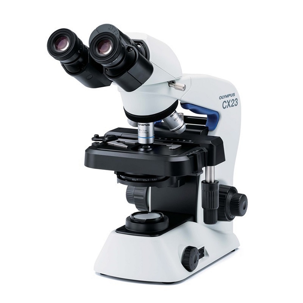
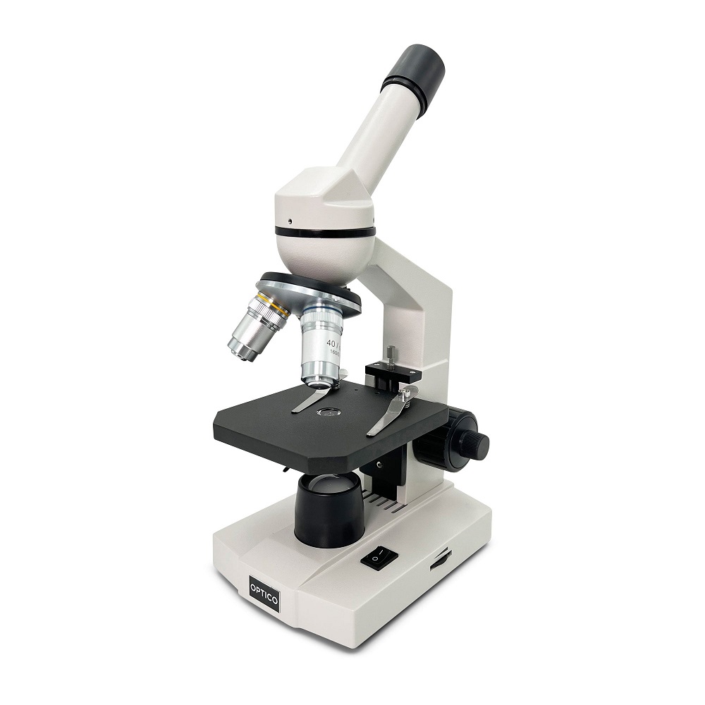
A light microscope is an optical instrument that uses visible light to magnify small samples. It typically consists of a series of lenses that focus light onto the specimen. The image formed is visible to the eye or can be captured using a camera. Light microscopes come in various types, including compound microscopes and stereo microscopes, each designed for specific imaging needs.
The magnification power of light microscopes usually ranges from 40x to 1000x. Although this may not be enough to view extremely small microorganisms, such as viruses, it is sufficient for examining cells and tissues. The clarity and detail offered by light microscopes make them vital instruments in many scientific fields.
History and Development
The history of the light microscope dates back to the 1600s when pioneers like Antonie van Leeuwenhoek first developed simple microscopes. Leeuwenhoek’s discoveries of single-celled organisms marked a significant milestone in microbiology. Over the years, improvements in lens design and optics have led to the development of compound microscopes, which use multiple lenses to enhance magnification and resolution.
With continued advancements in technology, modern light microscopes have become more powerful and versatile. They now incorporate features like digital imaging, fluorescence, and phase contrast. These enhancements have expanded the range of applications, making light microscopes a cornerstone in both research laboratories and educational settings.

Key Features of Light Microscopes
Optical Components
The quality of the optical components is critical to the performance of a light microscope. The two main optical components are the objective lenses and the eyepiece. Objective lenses are located near the specimen and are responsible for magnifying the sample. They come in various magnifications, usually ranging from 4x to 100x. The combination of objective and eyepiece lenses allows for a total magnification that can effectively enlarge the object being observed.
High-quality optics are essential for producing clear, crisp images. A well-constructed lens system minimizes optical aberrations and distortions, which can hinder the clarity of the image. The resolving power, or the ability to distinguish two closely spaced points, is an important specification that is determined by the lens quality.
Illumination Systems
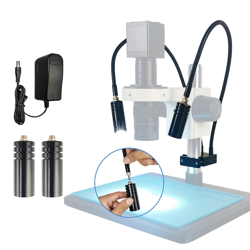
Proper lighting is another crucial aspect of the light microscope. Modern microscopes often employ various illumination techniques to enhance visibility and contrast. The most common type is brightfield illumination, where light passes directly through the specimen. However, this method may not be suitable for transparent or unstained samples.
To improve contrast in such samples, techniques like phase contrast and darkfield microscopy can be applied. Phase contrast microscopy allows for the observation of transparent specimens without staining, making it popular in biological research. Darkfield microscopy uses a special light path to illuminate the specimen from the side, resulting in a bright image on a dark background. These illumination systems enable scientists to visualize a wide range of specimen types effectively.
Types of Light Microscopes
Compound Microscopes
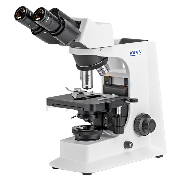
Compound microscopes are the most common type of light microscope. They are characterized by multiple objective lenses that are mounted on a rotating nosepiece. This design allows users to switch between different magnifications easily. Typical uses of compound microscopes include examining prepared slides of biological tissues, cells, and microorganisms.
Compound microscopes produce high-resolution images and are ideal for studying fine details in cellular structure. They are extensively used in laboratories, educational institutions, and clinics, making them indispensable tools in various fields, including biology, medicine, and pathology.
Stereo Microscopes
Stereo microscopes, also known as dissecting microscopes, provide a three-dimensional view of specimens. Unlike compound microscopes, stereo microscopes use two optical pathways to create a sense of depth, allowing for a more comprehensive view of larger specimens. They typically have lower magnification ranges (from 10x to 40x) compared to compound microscopes.
Stereo microscopes are commonly used in fields such as biology, botany, and electronics. They are helpful for examining small, solid objects, such as insects or circuit boards. The three-dimensional view simplifies tasks like dissection, assembly, and repair, making stereo microscopes valuable tools in both educational and research settings.

Applications in Biological Research
Cell Biology
In the realm of cell biology, light microscopes play a pivotal role in studying cellular structures and functions. Researchers use them to examine live cells in culture or analyze fixed samples. The ability to visualize organelles, cell membranes, and cytoskeletal components enhances our understanding of cell physiology.
Fluorescence microscopy, a technique that uses fluorescent dyes, is particularly valuable for observing specific cellular components. By tagging proteins or other molecules with fluorescent markers, researchers can study their localization and interactions within the cell. This approach has advanced our knowledge of cellular processes such as signaling pathways and disease mechanisms.
Pathology
Light microscopy is indispensable in the field of pathology, where it is used to diagnose diseases by examining tissue samples. Pathologists analyze stained sections of tissues to identify abnormal cellular structures or patterns that indicate disease. Techniques such as hematoxylin and eosin staining allow the visualization of various cell types and tissue architecture.
Furthermore, immunohistochemistry employs antibodies to detect specific proteins within tissues. This technique aids in identifying disease states, such as cancer, and determining the appropriate course of treatment. The ability to visualize tissue samples at the microscopic level is essential for accurate diagnoses and effective patient management.
Applications in Material Science
Study of Materials
In material science, light microscopes are employed to analyze the microstructure of materials. They help researchers investigate the properties and behaviors of metals, polymers, ceramics, and composites. The microscopic examination can reveal vital information about material composition, grain structure, and surface features.
Using polarized light microscopy, scientists can study crystalline materials with high precision. This technique allows for the identification of different phases and the characterization of mineral samples. Understanding the microstructural properties enhances the development of new materials and processes in various industries.
Quality Control
Light microscopy also plays a significant role in quality control across various manufacturing processes. Inspecting the microstructure of materials during production ensures that they meet the required specifications. Manufacturers can identify defects, inconsistencies, or impurities that may compromise product quality.
In industries such as electronics, light microscopy is used to examine components for defects and ensure reliability. By regularly testing and inspecting materials at the microscopic level, companies can maintain high-quality standards and improve product performance.

Educational Applications
Learning and Understanding
Light microscopes serve as valuable educational tools in schools and universities. They provide students with hands-on experience in biology, chemistry, and material science. Observing specimens under a microscope allows students to understand complex concepts, such as cell structure and tissue organization.
Using these microscopes during laboratory sessions enhances critical thinking and scientific skills. Students learn how to prepare samples, operate the microscope, and analyze images. This practical experience fosters curiosity and encourages exploration in the fields of science and technology.
Citizen Science Projects
In recent years, light microscopes have been utilized in citizen science projects that encourage public engagement in scientific research. These projects allow individuals from various backgrounds to contribute to scientific knowledge by analyzing local samples, such as soil, water, or plant specimens.
Participants can use light microscopes to observe microorganisms, identify species, and record their findings. This hands-on involvement democratizes science and promotes collaboration between scientists and the community. Such initiatives can significantly impact environmental monitoring and biodiversity studies.
Future Trends in Light Microscopy
Advancements in Technology
The field of light microscopy is continuously evolving, driven by advancements in technology. Innovations such as super-resolution microscopy and multi-photon microscopy have expanded the capabilities of traditional light microscopes. These techniques push the limits of resolution, allowing scientists to observe structures at previously unattainable levels.
As technology progresses, integrating artificial intelligence and machine learning into light microscopy will likely enhance image analysis. Automated software can optimize image capture, assist in identifying features, and analyze large datasets efficiently. These advancements can streamline research processes and lead to new discoveries in various scientific fields.
Integration with Other Techniques
The future of light microscopy also lies in its integration with other imaging techniques. Combining light microscopy with electron microscopy or X-ray imaging can provide complementary data about samples. This multi-modal approach can deepen our understanding of complex structures and interactions, paving the way for breakthroughs in material science and biology.
Furthermore, the development of portable light microscopes will increase accessibility for researchers and educators in remote areas. Such devices will make it easier to conduct field studies, enhance environmental monitoring, and promote scientific exploration. The potential for light microscopes to adapt and evolve ensures their continued relevance in scientific research.
The Enduring Importance of Light Microscopes
In conclusion, light microscopes are invaluable tools in various scientific disciplines. Their key features, including optical components, advanced illumination systems, and various types, enhance our ability to study the microscopic world. The applications of light microscopy span biology, material science, education, and public engagement, reflecting its versatility and importance.
As technology continues to advance, the future of light microscopy appears bright. The integration of innovative techniques and artificial intelligence will undoubtedly further enhance its capabilities. For researchers, educators, and students alike, light microscopes will continue to serve as gateways to discovery and understanding, enabling us to explore the intricate details of the world around us. Through the lens of a light microscope, we gain a deeper appreciation for biology, materials, and the scientific processes that shape our understanding of life.
