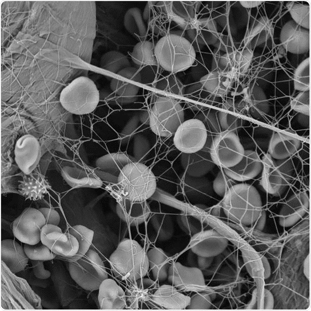
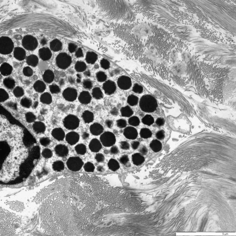
Electron microscopy has transformed the field of imaging, offering unparalleled resolution and detail at the nanoscale. As technology progresses, innovations in electron microscopy continue to push the boundaries of what can be observed and analyzed. This article explores the latest advancements in electron microscopy and their impact on various scientific fields. By understanding these key innovations, we can appreciate the future potential of this powerful imaging technique.
Understanding Electron Microscopy Basics
What is Electron Microscopy?
Electron microscope is a technique that uses electrons instead of light to form an image of a sample. This method provides much higher resolution than traditional optical microscopy, allowing scientists to observe structures at the atomic level. Electrons have much shorter wavelengths than visible light, which enables them to resolve smaller features in materials.
There are several types of electron microscopes, including transmission electron microscopes (TEM) and scanning electron microscopes (SEM). Each type has its specific applications and advantages. TEM allows scientists to view the internal structure of thin specimens, while SEM provides detailed images of surfaces. Understanding these basic principles is crucial for grasping the innovations that are shaping the future of this technology.
The Importance of Resolution and Contrast
Resolution and contrast are critical factors in microscopy. Resolution refers to the ability to distinguish two nearby points as separate entities, while contrast refers to the difference in brightness between an object and its background. In electron microscopy, achieving high resolution and contrast often involves complex techniques and sample preparation methods.
Innovations in electron microscope focus on enhancing both resolution and contrast. Improved detectors, advanced electron sources, and new imaging techniques contribute to these advancements. As researchers continually strive for better visualization, the significance of high-quality imaging becomes even more apparent, driving further developments in the field.

Advances in Resolution and Magnification
Super-Resolution Techniques
One of the most significant innovations in electron microscope is the development of super-resolution techniques. These methods allow scientists to achieve resolutions beyond the classical limits of electron microscopy. Researchers have developed techniques such as stimulated emission depletion (STED) and photo-activated localization microscopy (PALM), significantly advancing the field.
Through super-resolution, scientists can visualize nanoscale structures with remarkable detail. For instance, STED can achieve resolutions in the range of 20 nanometers, allowing researchers to explore intricate cellular structures and biomolecules. These breakthroughs have exciting implications for fields like biology and materials science, where understanding fine details is crucial.
Aberration-Corrected Electron Microscopy
Another essential advancement is the introduction of aberration-corrected electron microscopy. Aberrations are distortions that affect the quality of the image produced by an electron microscope. By using advanced optics and correction techniques, researchers can minimize these distortions and improve image clarity.
Aberration correction allows for achieving much higher resolutions than previously possible. This innovation is particularly important in materials science, where atomic-level analysis can reveal critical information about materials’ properties. As a result, aberration-corrected electron microscopes have become a valuable asset in research laboratories worldwide.
Enhanced Imaging Techniques
High-Throughput Imaging
High-throughput imaging techniques are rapidly emerging in electron microscopy. These methods enable researchers to analyze multiple samples quickly, increasing productivity in research settings. Utilizing automated systems and advanced software, scientists can acquire large datasets efficiently.
High-throughput imaging is especially beneficial for large-scale studies in materials science and biology. For example, researchers can screen vast libraries of nanomaterials to identify promising candidates for specific applications. The ability to gather extensive image data quickly offers significant advantages for developing new materials and therapies.
Cryo-Electron Microscopy
Cryo-electron microscopy (cryo-EM) has gained significant attention in recent years. It involves cooling samples to extremely low temperatures, preserving their native structure. Cryo-EM has become a powerful tool for visualizing biological molecules, such as proteins and viruses, in their functional states.
The innovation of cryo-EM allows scientists to study macromolecules without the need for crystallization or extensive sample preparation. This has led to breakthroughs in understanding biological processes at the molecular level. Notably, cryo-EM played a critical role in elucidating the structures of several important proteins, including those related to diseases.

Applications Across Scientific Disciplines
Materials Science
In materials science, electron microscopy is essential for characterizing materials at the nanoscale. This involves studying the microstructure, crystallography, and defects in materials. Innovations in electron microscopy, such as enhanced imaging techniques and advanced resolution, provide deeper insights into material properties.
Understanding materials at the atomic level is crucial for developing new technologies, including semiconductors, superconductors, and nanomaterials. The ability to visualize the structure of these materials helps researchers optimize their performance in various applications. Thus, advances in electron microscopy directly contribute to progress in materials science.
Biology and Medicine
Electron microscopy has revolutionized biological research, particularly in the field of cell biology. The ability to visualize cellular structures at high resolution enables researchers to study the intricate details of organelles, membranes, and biomolecules. Techniques like cryo-EM have propelled biological imaging into new territories, allowing scientists to examine proteins and viruses in their native states.
In medicine, electron microscopy has been vital for diagnosing diseases and understanding pathological processes. Researchers can examine tissue samples to identify abnormalities and understand disease mechanisms. With innovations in imaging and resolution, electron microscopy continues to play a critical role in advancing medical knowledge and patient care.
Integration with Multi-Modal Techniques
Combining Electron Microscopy with Other Methods
The integration of electron microscopy with other imaging techniques has become increasingly common. Combining methods enhances the overall understanding of complex samples. For example, pairing electron microscopy with atomic force microscopy (AFM) or X-ray diffraction can provide complementary information about material properties.
By leveraging the strengths of multiple imaging techniques, researchers can gain a more comprehensive understanding of their samples. The convergence of different modalities allows for detailed structural analysis and insights into the functional properties of materials or biological systems.
Advancements in Software and Image Analysis
Innovations in software development have also transformed electron microscopy. Advanced image processing and analysis tools enable scientists to extract valuable information from large datasets. Machine learning and artificial intelligence algorithms can help automate the analysis process, recognizing patterns and features in images more efficiently.
These advancements assist researchers in interpreting complex data sets generated by high-throughput imaging. As the volume of data increases, automated analysis will become increasingly vital. This combination of imaging techniques and sophisticated software tools will enhance the capabilities of electron microscopy even further.

Challenges and Considerations
Cost and Accessibility
Despite the significant advancements in electron microscopy, challenges remain, particularly concerning costs. High-resolution electron microscopes, especially those with advanced features, can be prohibitively expensive. The cost of maintenance and operation can also be substantial, limiting access for some institutions, particularly smaller research laboratories.
Efforts are underway to develop more affordable and user-friendly electron microscopy systems. Making these technologies more accessible would democratize research and enable a wider range of scientists to utilize electron microscopy in their work.
Sample Preparation
Sample preparation continues to pose challenges in electron microscope. Properly preparing samples is essential for obtaining high-quality images. However, preparing biological samples often requires extensive procedures that can introduce artifacts or alter the samples’ properties.
Innovations in sample preparation methods, including cryo-fixation and automated systems, aim to minimize these issues. Researchers are actively exploring new techniques to improve sample preparation, ensuring that the integrity of the specimens is preserved. Continued advancement in this area is critical for maximizing the potential of electron microscopy.
The Future of Electron Microscopy
Promising Directions
Looking ahead, the future of electron microscope is promising. Continued advancements in technology, resolution, and methodology will further enhance its capabilities. As new innovations emerge, electron microscopy will play an increasingly prominent role in diverse scientific fields. Fields such as nanotechnology and synthetic biology will particularly benefit from enhanced imaging techniques.
Future developments may also focus on enhancing usability. Streamlining workflows and simplifying operation will attract more researchers to utilize electron microscopy. User-friendly interfaces and automated systems can lower the barriers to entry, driving wider adoption across laboratories and educational institutions.
A Bright Future
In conclusion, electron microscope remains an essential tool in scientific research, offering unprecedented imaging capabilities. Innovations in resolution, imaging techniques, and integration with other technologies will continue to propel the field forward. As scientists explore new frontiers, electron microscopy will provide insights into the nanoscale world, enabling discoveries that shape our understanding of materials, biology, and the universe.
The evolution of electron microscope over the years illustrates the adaptability and growth of this powerful technique. Its future is bright, filled with opportunities and advancements that will change how we perceive and understand the intricate details of the world around us. By investing in technology and fostering collaboration within the scientific community, the potential for electron microscopy is limitless.
