Introduction to Pineapple Microscopy
The study of pineapples under a microscope opens up a fascinating world. Pineapple microscopy examines the fruit’s intricate details that we can’t see with the naked eye. It reveals the cellular structure, enzyme activity, and fiber composition. This microscopic analysis helps us understand why pineapples have unique textures and flavors.
When looking at a pineapple under the microscope, we dive deep into its biology. We see cells working together to form this tropical treat. Our investigation can also uncover pests and diseases at an early stage. This can prevent crop damage. Moreover, it tells us about the fruit’s nutritional makeup in minute detail. Understanding how to view a pineapple under microscope is crucial for this exploration. Advanced techniques and tools are necessary.
Scientists and researchers use pineapple microscopy to study growth, ripening, and spoilage. They work to enhance our knowledge and improve pineapple crops. With this microscopic insight, the future of pineapple research looks bright. We can develop new ways to protect and boost the health benefits of pineapples. Let’s explore the cellular secrets of this popular fruit together.

The Pineapple’s Cellular Structure
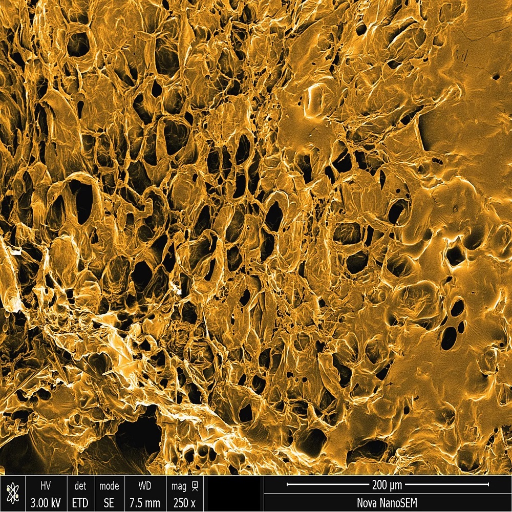
The cellular structure of a pineapple is as unique as its tangy taste. When observing a pineapple under microscope, we unveil a network of cells bound tightly in an organized pattern. This structure is key to the pineapple’s durability and juicy texture.
Each cell in this pattern stores nutrients and water. This helps the fruit stay hydrated and nutrient-rich. The pineapple’s cells also have strong walls. These walls give the fruit its firmness. They are why a pineapple does not lose its shape easily, even when ripe.
Interestingly, the pineapple’s cells differ in size and shape. This variety depends on their position within the fruit. Cells near the core are often more elongated. This supports the pineapple’s structural integrity. Meanwhile, those towards the outer flesh are rounder and packed with juice. These cells contribute to the sweet and refreshing flavor we love.
Chlorophyll is another component found within the pineapple’s cells. It gives unripe pineapples their green hue. As the pineapple ripens, the chlorophyll breaks down. This is when the fruit turns to its golden-yellow color that signals readiness to eat.
Viewing the cellular structure can also aid in understanding ripening stages. Changes in cell size and wall density indicate how far along the fruit is in its life cycle. This microscopic insight is crucial for farmers and researchers alike.
In summary, the pineapple’s microscopic cellular structure is not just a biological marvel. It is also central to its taste, texture, and nutritional value. Understanding this on a micro level is vital in the study of pineapples.
Visualizing Pineapple Enzymes at the Microscopic Level
When we place a pineapple under microscope, enzymes become visible. These enzymes are vital for the fruit’s life. They break down proteins and aid in ripening. Studying them helps us understand how pineapples become juicy and sweet.
One key enzyme visible at this level is bromelain. Bromelain is known for its ability to tenderize meat. This is because it breaks down protein structures. Under the microscope, we can see bromelain’s activity. It’s fascinating to watch it work at the cellular level.
Another set of enzymes helps defend the fruit from pests. These enzymes become active when the fruit is damaged. This is pineapple’s natural defense mechanism. It’s an impressive process to see under the microscope.
Enzymes also play a role in the pineapple’s color change. As mentioned, chlorophyll breaks down when the fruit ripens. Enzymes facilitate this breakdown. Viewers can see the gradual color shift under the microscope.
Analyzing pineapple enzymes microscopically does more than satisfy curiosity. It has practical uses too. For example, it can help improve food processing techniques. It can also lead to the development of medicinal uses for bromelain.
Overall, enzymes in pineapples are a window into the fruit’s inner workings. They are fascinating to study and have important roles. With a microscope, we uncover their part in making the pineapple a unique fruit.

The Role of Microscopic Fibers in Pineapple Texture
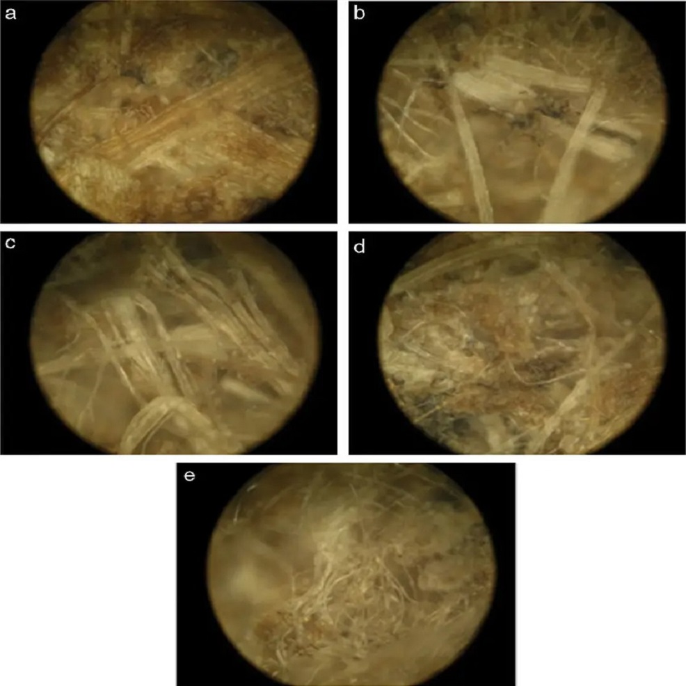
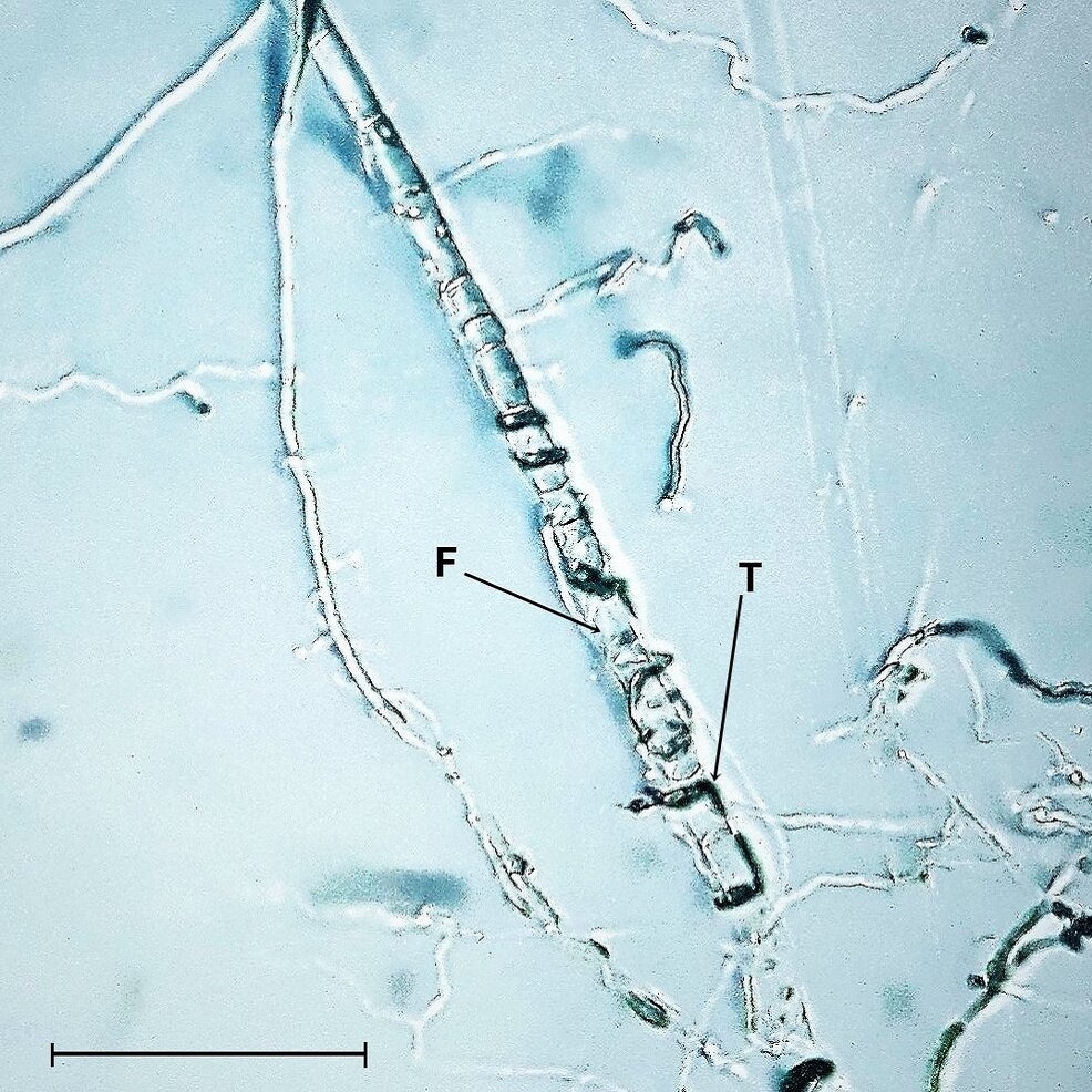
The texture of a pineapple is crucial to its appeal. Microscopic fibers play a significant role in defining it. Under a microscope, we can see a vast network of fibers threading through the pineapple. These fibers are responsible for the fruit’s structural integrity and its signature chewiness.
Pineapple fibers, mainly composed of cellulose, are tightly packed. This packing contributes to the firmness of the pineapple. It’s what makes the pineapple withstand external pressure without collapsing. When you bite into a pineapple, it’s the strength of these fibers you’re feeling.
In the core, fibers are more densely arranged. This is why the core feels tougher to eat compared to the softer flesh. The softer outer flesh has fibers spaced further apart. This spacing allows for more juice storage, resulting in that juicy burst of flavor we enjoy.
Microscopic examination of pineapple fibers also reveals their link to ripeness. As pineapples ripen, the fibers become more flexible. This flexibility gives ripe pineapples their pleasant mouthfeel. However, overripe fruits may have too soft fibers, leading to mushiness.
Farmers and food scientists value this knowledge highly. Understanding fiber distribution and density helps in selecting the best pineapples for consumption. It also aids in determining the optimal ripening time for harvesting. Knowledge of fiber structure can even influence the development of new pineapple varieties. These varieties could have desired textures and structural properties.
Overall, the intricate network of microscopic fibers is key to the pineapple’s texture. It affects everything from firmness to juiciness, influencing both taste and customer satisfaction. Viewing these fibers under a microscope adds depth to our understanding of this popular fruit.
Microscopic Pests and Diseases Affecting Pineapples
When we examine a pineapple under microscope, we sometimes find unwanted guests. Microscopic pests and diseases can lurk within the fruit. Spotting them early can prevent them from spoiling the crop. Under high magnification, researchers can see tiny insects and fungal spores. These can harm the plant’s health and the fruit’s quality.
Common pests include the pineapple mealybug and the pineapple mite. These tiny creatures feed on the pineapple’s sap. They weaken the plant and can spread viruses. Under the microscope, their presence becomes clear. This helps farmers take action to protect their crops.
Diseases also threaten pineapples. One example is the pineapple heart rot. This disease causes the fruit’s core to rot. Microscopic fungi, like Phytophthora cinnamomi, lead to this condition. Using a microscope, experts can spot the early signs of fungal growth. They can then use the right treatments to stop the spread.
Another disease visible under the microscope is Fusarium wilt. This fungus attacks the pineapple plant’s vascular system. It can cause leaves to wilt and fruits to develop unevenly. Early detection through microscopy can save a whole plantation.
In conclusion, studying pineapples under a microscope is not just about beauty. It is a critical practice to identify and manage pests and diseases. Timely action based on microscopic observation safeguards the health of pineapple crops.
Nutrition Under the Microscope: The Composition of a Pineapple
When we examine a pineapple under microscope, its nutritional composition comes to light. A close-up view reveals vitamins, minerals, and sugars that make the pineapple a healthy snack. Here, we’ll break down what these microscopic discoveries tell us about pineapple nutrition.
First, we find the building blocks of health: vitamins. Pineapples are rich in vitamin C, essential for immunity and skin health. A microscope shows us the dense clusters of vitamin C molecules within its flesh. These molecules protect the fruit’s cells, much like they protect ours.
Minerals are next. Magnesium and potassium appear under the lens. These minerals are crucial for heart function and muscle contraction. Microscopic examination shows their dispersion throughout the fruit, contributing to its overall nutrient profile.
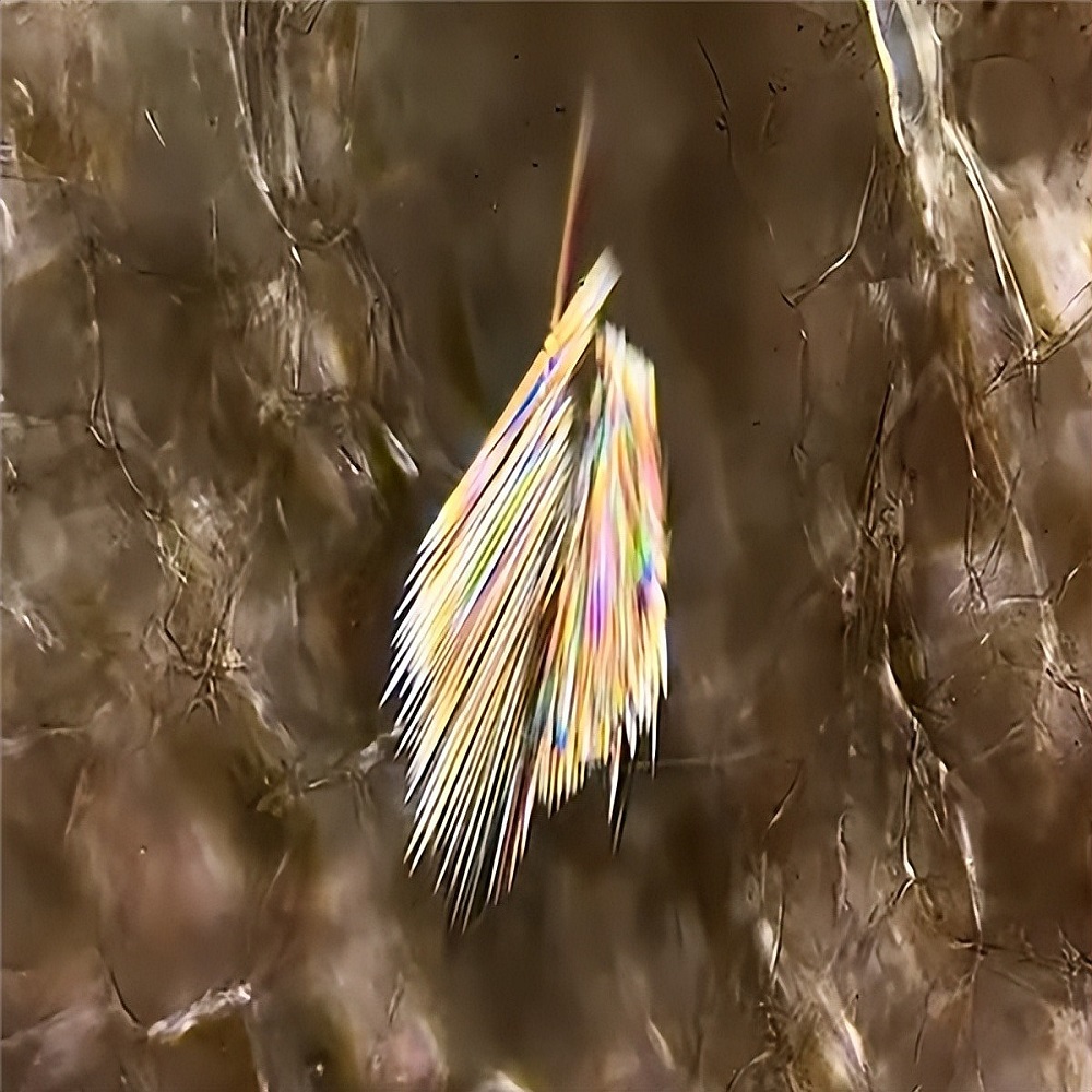
Sugars like glucose, fructose, and sucrose give pineapples their sweetness. Under high magnification, these sugars emerge as crystalline structures. Pineapples are a natural source of these sugars, providing us with quick energy.
Microscopic analysis also detects dietary fibers. These fibers are vital for digestion and fullness. The closely woven fibers form networks that help us understand how pineapples aid in gut health.
Pineapple’s water content is also high. Up close, we see how water is stored within the fruit’s cells. This hydration is a key part of the fruit’s succulence. It also makes pineapples a refreshing choice in the diet.
Finally, under the microscope, we may find trace elements. These include iron and zinc, albeit in smaller quantities. These elements play parts in various body functions, including oxygen transport and immune responses.
In conclusion, viewing a pineapple under microscope uncovers a wealth of nutritional benefits. Each vitamin, mineral, and sugar plays a role in making the pineapple not just a treat, but a nutritious choice as well.

Techniques and Tools: How to View a Pineapple Under the Microscope
For those curious about viewing a pineapple under microscope, certain tools and techniques are essential. Firstly, a high-quality microscope with the ability to magnify objects at least a hundred times is necessary. To prepare the pineapple for microscopic examination, thin slices which allow light to pass through are needed. These slices should be cut using a sharp blade or a special tool like a microtome to ensure precision and consistent thickness.
A glass slide and cover slip are staples in this process. They hold the pineapple sample in place. Staining techniques might also be used to enhance visibility of certain cells or enzymes. These stains attach to specific components, making them stand out. It’s important to choose stains that target the elements you wish to examine – like chlorophyll or bromelain.
While working under the microscope, proper lighting is crucial. Many microscopes come with built-in lights. Yet, sometimes additional light sources are needed to illuminate the sample. Digital microscopes are also an option. They allow researchers to see and capture images on a computer screen. This makes it easier to share and analyse the findings.
In conclusion, viewing a pineapple under microscope calls for the right set of tools and techniques. From cutting thin samples to applying stains, and from using a powerful microscope to ensuring good lighting, each step is key. With this approach, we can visualize the unseen beauty and complexity of pineapples.
The Future of Pineapple Research and Microscopy
The future of pineapple research through microscopic analysis is promising. By placing a pineapple under microscope, scientists can advance in many areas. They can improve crop yields, enhance fruit quality, and develop better disease-resistant varieties. Here are the ways in which pineapple microscopy is shaping the future:
- Crop Improvement: Researchers study cellular patterns to create stronger pineapples. These could resist pests and harsh weather better.
- Disease Management: Early detection of microscopic fungi and pests can prevent outbreaks. This saves large-scale crops and ensures healthy fruit production.
- Quality Control: Through enzyme visualization, scientists work on optimal ripening processes. This could mean sweeter, more flavorful pineapples for consumers.
- Nutritional Enhancement: By analyzing the nutrients in pineapples, new varieties might be bred. These varieties could have higher vitamins and minerals.
- Innovative Techniques: The use of digital microscopes and advanced staining methods will grow. These tools make research easier and more accurate.
In the years to come, researchers aim to team up with farmers. They want to apply microscopic insights directly to agriculture. They also plan to use data to predict and tackle diseases before they spread.
Finally, educational initiatives could play a role. Schools and universities may use pineapple microscopy in teaching. Students could learn about plant biology in a vivid and hands-on way.
To sum up, the world of pineapple microscopy is evolving. It is opening new doors for research, health, and education. The tiny details seen under the microscope have big impacts on pineapple farming and consumption. The possibilities are as exciting as they are endless.
