Introduction to Hair Analysis
Hair analysis, using a Microscope, provides magnified details of hair. It is more than a diagnostic tool. Hair under microscope reveals growth patterns, structure, and even lifestyle markers. Experts use this method in many fields.
For example, forensic scientists analyze hair for criminal investigations. Health professionals may study hair to diagnose disorders. Also, cosmetic companies test hair for product development. Even environmental monitoring can involve hair analysis. All these applications rely on visual detail enhanced significantly under high magnification.
An expert may look at the hair cuticle, cortex, and medulla. Each part tells a different story. Damage, drug use, or dietary habits can become visible. These findings can guide various professional decisions. With a microscope, specifics of hair health and history become clear.
Evaluating hair under the microscope covers more than identification. It can also gauge hair care effects. High-quality microscopes show even the subtlest changes. With careful analysis, a professional can recommend changes for better hair health.
The process is intricate and requires proper technique. Preparing a hair sample involves careful collection and handling. Next, mounting the sample onto a slide follows. Staining is sometimes used to highlight features. Finally, microscopy can begin. Since hair structures are small, attention to detail is key. Magnification comes in different levels, offering different insights.
In the following sections, we’ll dive deeper into the types of hair viewed under the microscope, their structure, and what they tell us about health indicators, forensic science, disorders, and technological advancements.
Types of Hair Under the Microscope
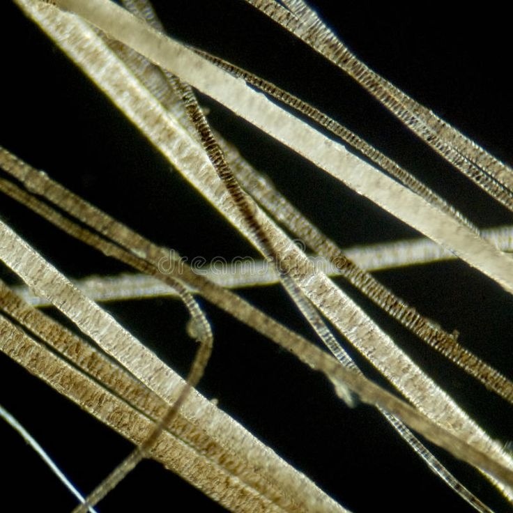
When we place hair under a microscope, we uncover a world of diversity. Not all hair is the same and understanding the different types can be crucial for experts in various fields. Here’s a detailed look at the various types of hair one might encounter under microscopic examination:
The Terminal Hair
This is the thick, long hair found on the scalp, face in men, and body in both sexes. Under the microscope, terminal hair reveals a robust medulla, a supportive cortex, and a protective cuticle layer. This type of hair often shows the most evidence of styling damage or environmental exposure.
Vellus Hair
Characterized by its fineness and short length, vellus hair is often referred to as ‘peach fuzz.’ Located all over the body, vellus hair is normally devoid of pigment and helps with temperature regulation. Microscopy of vellus hair displays a lack of medulla, signifying its delicate nature.
Lanugo Hair
This is a fine, soft hair, typically found on newborns. It usually disappears shortly after birth. Lanugo hair under a microscope appears wispy and is an indicator of the gestational stage in infants.
Beard Hair
Beard hair, seen under magnification, shows a unique coarseness compared to scalp hair. It has a circular cross-section and may show varying degrees of curliness, which can influence beard patterns and styling.
Each hair type, when magnified, reveals intricate details about the individual’s health, lifestyle habits, and even genetic background. Hair under microscope examination enables a meticulous study of these differences and contributes to a better understanding of human biology and health.
The Structure of a Hair Strand
When examining hair under microscope, we observe distinct features. Each hair strand is unique. The strand has a complex structure with three main components. These are the cuticle, cortex, and medulla.
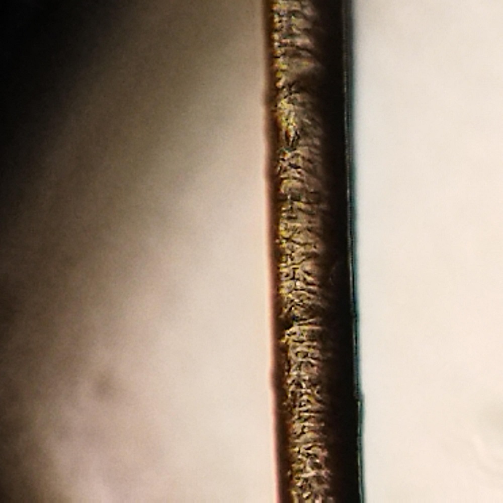
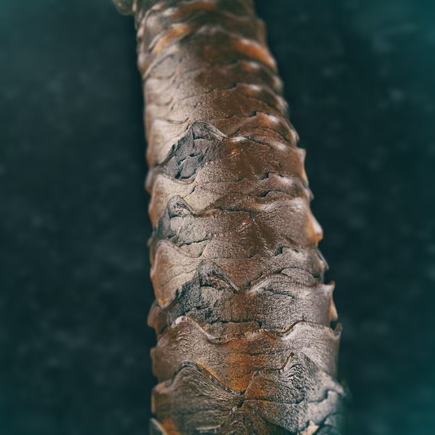
Cuticle
The cuticle is the outermost layer. It’s made of overlapping scales. These protect the inner layers. It also gives hair its shine. Damage to the cuticle can lead to dull, brittle hair.
Cortex
Below the cuticle lies the cortex. It contains long keratin fibers. It’s responsible for strength and elasticity. The cortex also holds melanin. This gives hair its color.
Medulla
The medulla is the core. It’s a soft, central column. In some hair types, it may be absent. Its purpose is still not fully understood.
The microscope reveals how these structures interact. It shows signs of health and damage too. By analyzing these components, experts learn a lot. They understand more about personal health and habits. Harsh chemicals or heat can damage the hair. Poor nutrition shows in the hair structure as well. This is why studying hair under microscope is vital. It gives accurate insights into our health and lifestyle.
Differentiating Between Animal and Human Hair
Under the lens of a microscope, distinguishing between animal and human hair becomes a precise science. Despite both having the same basic structure—cuticle, cortex, and medulla—there are marked differences in the microscopic characteristics.
Cuticle Patterns
Animal hair often displays more varied and distinct cuticle patterns compared to human hair. These reflect species-specific adaptations.
Medulla Structure
The medulla in animal hair is usually more prominent and displays diverse patterns. In contrast, human hair has a less pronounced medulla, sometimes entirely absent.
Hair Shaft Diameter
Animal hair shafts vary greatly in diameter, sometimes even within the same species. Human hair tends to have a more uniform and narrower diameter.
Pigmentation
Pigment granules in animal hair are typically more densely packed and can show a more uneven distribution than in human hair. Human hair pigmentation is generally more homogenous.
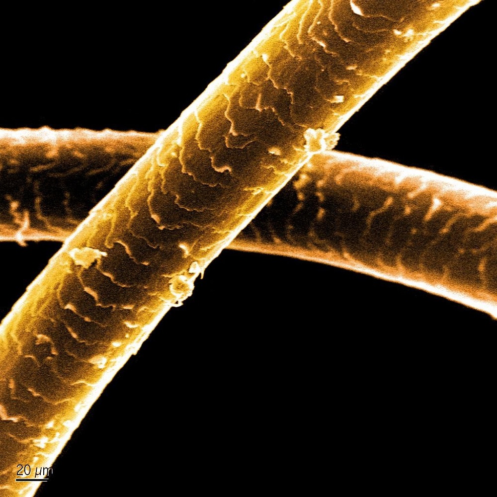
Scale Structure
The scales on animal hairs are often more petal-like and imbricate differently. Human hair scales are less pronounced and tend to have a smoother look.
These features provide vital clues in forensics and wildlife studies. They help experts in pinpointing species, understanding habitats, and solving crimes. Hair under microscope study exposes these differences with clarity, offering a window into the distinct worlds of human and animal hair.
Hair Health Indicators: What Microscopy Can Reveal
Using a microscope to observe hair can tell us much about our health. By looking at hair under microscope, experts see clues that suggest our well-being. Here, we will explore what indicators can be uncovered through hair microscopy.
Firstly, damage to hair is readily apparent when magnified. Broken cuticles and split ends can show rough treatment or neglect. Next, hydration levels become obvious. Healthy hair will appear smooth and intact, while dry hair will look brittle and frayed.
Also, our lifestyle choices may reflect in our hair. For instance, repeated chemical treatments reveal themselves in damaged cortex fibers. Furthermore, the presence of drugs or toxins can often be detected in the medulla. Even dietary patterns can influence hair quality, visible under high magnification.
Moreover, hair can reflect general health issues. For example, dull, lifeless hair might indicate a lack of nutrients or underlying health conditions. On the other hand, shiny, resilient hair may suggest good health and care.
By using a microscope to look at hair, professionals can offer tailored advice for improving hair health. This microscopic examination is a tool for both diagnosis and monitoring. It helps us see the links between our habits, health, and the appearance of our hair. Each strand tells a story, unveiling secrets of our inner health and lifestyle choices.
The Role of Hair Analysis in Forensic Science
Hair under microscope examination plays a critical role in forensic science. It provides irrefutable clues in solving crimes. As each hair type has distinct microscopic features, they can be powerful evidence. Forensic experts analyze these features to draw conclusions about a person’s actions or whereabouts.
Identifying Individuals
Through microscopic analysis, unique patterns in hair can help identify people. Personal hair characteristics like color, thickness, and cuticle patterns are key. No two individuals have identical hair profiles, enabling personal identification.
Linking Suspects to Crime Scenes
Microscopy can tie suspects to crime scenes. Finding a hair sample at a crime site can link a person to that place. Forensic analysts compare samples from suspects to those found on-site.
Determining Timeframes
Hair grows at a known rate. This growth rate can help estimate when a hair was last cut or dyed. Such timelines are crucial in crime investigations.
Unearthing Lifestyle Choices
Hair under microscope can reveal lifestyle habits, like drug use. These details can support other evidence in legal cases. Microscopic hair analysis highlights these habits clearly and accurately.
Forensic hair analysis under a microscope is an essential tool. It is indispensable in criminal investigations. It uncovers hidden truths and reinforces the accuracy of forensic evidence. These microscopic insights make the invisible, visible; crucial in the pursuit of justice.
Hair and Scalp Disorders Observed Microscopically
When experts put hair under a microscope, they can spot various health problems. Scalp and hair disorders often show up with clear signs when magnified. Let’s look at what disorders can be seen and what they may indicate.
Scalp Issues
Problems like dandruff, psoriasis, and folliculitis are common scalp disorders. These conditions can cause flaking, redness, and inflammation. Under a microscope, signs like abnormal skin scaling and changes in hair follicles become evident.
Hair Shaft Abnormalities
Abnormal hair shafts may point to genetic disorders or damage from treatments. Microscopy can show kinks, thinning, and brittleness. These often result in weak hair that breaks easily.
Pattern Baldness
Pattern baldness, or androgenetic alopecia, is recognizable by the miniaturization of hair follicles. Microscopically, one can observe a gradual decrease in hair shaft diameter which leads to thinning hair.
Infections
Fungal infections such as ringworm show up as changes in the hair cuticle and shaft. Bacterial infections can damage the hair cuticle and cortex, seen under high magnification.
By examining hair under microscope, experts can identify these disorders early on. Timely diagnosis and treatment can improve scalp health and hair condition.
Advancements in Microscopic Hair Analysis Technology
In the quest to understand hair under a microscope better, technology has come a long way. Modern advancements in microscopic hair analysis have enhanced the capabilities of experts in various fields, providing more precise and detailed observations.
Improved Magnification Techniques
Advancements in magnification have allowed for higher resolution imagery of hair strands. With stronger zoom capabilities, scientists can see fine details that were once blurry or invisible. This helps in identifying minute signs of damage or health.
Digital Microscopy Integration
Digital microscopes now replace traditional ones in many cases. They offer clearer images and the ability to document and share findings easily. With this technology, hair analysis becomes more efficient and collaborative.
Software for Hair Analysis
Specialized software can now assist in analyzing hair structures more quickly. Programs can measure hair features, compare patterns, and even predict certain health conditions based on hair traits.
Automation in Sample Preparation
Preparing hair samples for microscope analysis can be tedious. However, new tools now automate this process, reducing human error and saving time. This step ensures more consistent and reliable observations.
Non-Invasive Techniques
Some of the latest techniques for examining hair don’t even require cutting a strand. Non-invasive methods safely analyze hair on the scalp, keeping the sample intact.
These advancements make hair under microscope analysis more powerful than ever. It remains an essential method for understanding hair-related health, forensics, and cosmetic research. As technology progresses, we can expect even greater accuracy and insight from microscopic hair studies.