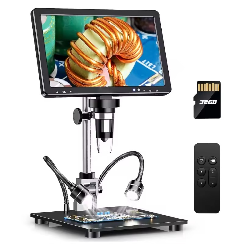
Introduction to Electron Microscopy
Electron microscopy has revolutionized how we see the world at the micro level. Using a beam of electrons, this powerful tool can reveal structures far too small for optical microscopes to capture. The difference lies in the wavelength of electrons, which is much shorter than that of light, allowing for much higher resolution images. Electron microscope images have thus become crucial in fields like materials science, biology, and nanotechnology, where understanding minute details is essential.
Typically, preparing a sample for viewing under an electron microscope is a meticulous process. It may involve careful slicing, drying, and even staining to provide the necessary contrast. Once ready, the sample is placed into the electron microscope’s vacuum chamber. Here, it is bombarded with electrons. The electrons interact with the sample’s atoms, creating various signals. These signals are then detected and transformed into detailed images.
Electron microscopy is also notable for its depth of field. This feature allows researchers to obtain a clear, focused view throughout the thickness of the sample. With this capability, it’s possible to explore the intricate topography of surfaces and the fine inner structures of specimens with astonishing clarity.
Overall, electron microscopy stands as a gateway into the unseen world, offering a unique glimpse into the fabric of materials and living organisms at the atomic level. Its ability to magnify and resolve details invisible to the naked eye has been invaluable in scientific discovery and innovation.

Key Components of an Electron Microscope
An electron microscope relies on several key components to produce highly detailed images. These key parts are crucial for its operation and performance. Let’s dive into these components and understand their roles.
Electron Source
The electron source, often a heated tungsten filament, generates the electrons needed for imaging. It acts much like a light bulb in an optical microscope, but it emits electrons instead of photons.
Electromagnetic Lenses
These lenses focus the electron beam onto the sample. While optical microscopes use glass lenses, electron microscopes use a series of electromagnetic fields to control the electron path.
Vacuum System
Maintaining a vacuum is essential as electrons are easily scattered by air molecules. The vacuum system removes air and other particles, allowing the electrons to reach the sample without interference.
Sample Holder
A precise sample holder secures the specimen in place inside the microscope. The sample must remain still to achieve clear images.
Detectors
After the electron beam interacts with the sample, detectors capture the resulting signals. These signals are then converted into the electron microscope images we analyze.
Display System
Finally, a display system, usually a computer monitor, visualizes the images generated from the detected signals. This allows researchers to examine the sample details.
Each of these components works in harmony to give a detailed view of tiny structures. By understanding how they contribute to creating electron microscope images, we can appreciate the complexity and precision of this scientific tool.
The Process of Capturing Images with an Electron Microscope
Understanding how electron microscope images are captured is fascinating. It involves a series of precise steps that require careful preparation and handling of the specimen. Here, we’ll outline this process, shedding light on how scientists and researchers use electron microscopes to get a closer look at the microscopic world.
First, a scientist prepares the specimen. This may include slicing it thinly, drying it out, or adding a contrast agent. The goal is to make the sample suitable for electron beams. Next, the specimen is placed on a grid or holder. This part must be handled with care to avoid damage.
The grid with the sample then goes into the electron microscope’s vacuum chamber. Air and moisture could scatter the electrons, and the vacuum helps prevent that. Once inside, the microscope aligns the electron beam with the sample using electromagnetic lenses.
The electron source sends out a stream of electrons. The beam is focused and directed onto the specimen. As the electrons hit the sample, they are either absorbed or scattered. Detectors inside the microscope capture these interactions.
Lastly, the signals detected by the microscope are converted. A digital image is formed on the display system for researchers to analyze. These images are highly detailed, providing valuable insights into the sample’s structure.
Every electron microscope image is a result of this meticulous process. Through it, we can explore microscopic dimensions with stunning clarity and detail.

Applications of Electron Microscopy in Various Fields
Electron microscopy has widespread use across various research disciplines. Each field leverages the power of electron microscope images to uncover new information and insights. Here’s a look at some notable applications:
- Materials Science: Researchers study the atomic structure of materials. They see how atoms arrange themselves and how this affects the material’s properties.
- Biology: Scientists explore cell structures, viruses, and bacteria. They gain a deeper understanding of life at the cellular level.
- Medicine: Medical professionals diagnose diseases by examining tissue samples. They look for abnormalities that the naked eye can’t see.
- Nanotechnology: Experts design and analyze nano-scale devices. They ensure these tiny gadgets are built correctly.
- Geology: Geologists examine minerals and rocks. This reveals their history and helps in predicting future changes.
- Forensic Science: Forensic experts use it to analyze crime scene evidence. Tiny details can often reveal crucial clues.
- Semiconductors: The tech industry inspects chips and circuitry. They find faults and improve designs for better electronics.
Electron microscope images continue to provide invaluable insights. They push the boundaries of what we know in science and technology.
Comparing Electron and Optical Microscopes
When examining the microscopic world, two types of microscopes come into play: electron and optical microscopes. These instruments share the common goal of magnification but differ fundamentally in technology and capability.
Optical microscopes, the more traditional form, use visible light to illuminate samples. They have glass lenses that bend light waves to enlarge the image of an object. Because they operate within the light spectrum, their resolution is limited by the wavelength of light. Typically, optical microscopes can magnify up to 1,000 to 2,000 times while retaining clarity.
Electron microscopes, on the other hand, employ a beam of electrons for illumination. Since electrons have a much shorter wavelength than light, electron microscopes surpass the resolution limit of optical ones. They can magnify objects up to two million times, revealing structures at the nanometer scale.
Another key difference lies in the medium used. Optical microscopes can examine samples in air or water, making them suitable for live specimens. Electron microscopes require a vacuum to operate, meaning samples must be dead and often coated or stained to withstand the vacuum and electron beam.
Depth of field is another area where the two microscopes diverge. Electron microscopes provide a greater depth of field, allowing for a three-dimensional examination of a specimen’s topography and composition. Optical microscopes have a shallower depth of field, which can limit the perception of three-dimensional structures.
Lastly, usability and cost are important considerations. Optical microscopes are common in schools and basic research due to their simplicity and affordability. Electron microscopes are more complex, require trained operators, and are significantly more expensive.
In summary, electron microscopes offer higher resolution and greater depth of field but at a higher cost and with more sample preparation. Optical microscopes are more accessible, easier to use, but limited in magnification and resolution.

Electron Microscopy Techniques: SEM and TEM Explained
When delving into electron microscope images, two advanced techniques stand out: SEM (Scanning Electron Microscopy) and TEM (Transmission Electron Microscopy). Both methods are critical in the world of electron microscopy, offering unique ways to examine samples.
Scanning Electron Microscopy (SEM)
SEM involves scanning a sample with a focused electron beam. Here’s how it works:
- Electrons sweep across the surface.
- They cause other electrons on the sample to eject.
- Detectors collect these secondary electrons.
- The signals create detailed surface images.
SEM is famed for its impressive three-dimensional images. It helps us understand surface textures and compositions with stunning clarity.
Transmission Electron Microscopy (TEM)
TEM, in contrast, transmits electrons through a sample. Here’s the process:
- A beam of electrons passes through a thin sample.
- The sample partly absorbs or scatters the electrons.
- Detectors capture the electrons that emerge.
- The signals form a detailed image of the sample’s inner structure.
TEM is ideal for viewing the internal makeup of cells, molecules, or materials at the atomic scale. It can show us features that are not visible with SEM.
Both SEM and TEM are powerful tools that provide electron microscope images of high resolution. While SEM gives us a detailed view of the sample’s surface, TEM offers an in-depth look at the internal structures. Together, they offer complementary insights that are invaluable across various scientific fields.
Advancements in Electron Microscopy Technology
Recent advancements in electron microscopy technology have greatly enhanced the ability to capture electron microscope images at unprecedented resolutions. These innovations have come from various aspects of the technology, each contributing to the enriched detail and clarity of the images we can now obtain.
Enhanced Electron Sources: New electron sources, like field emission guns (FEG), provide a brighter, more coherent electron beam. This sharpens image resolution significantly.
Improved Detectors: Detectors with higher sensitivity allow for better capture of the electrons that interact with the sample. This leads to crisper, more detailed images.
Advances in Digital Imaging: The transition to digital imaging systems has streamlined the process of capturing and analyzing electron microscope images. Higher resolution sensors and better data processing software aid in deciphering intricate details.
Development of In-situ Capabilities: Electron microscopes can now observe samples under various conditions – like different temperatures or mechanical stress – in real-time. This aids in understanding how materials behave in different environments.
Progress in Sample Preparation: New methods in sample preparation preserve samples better and enhance the contrast of the images. This allows for a clearer view of biological specimens, in particular.
Corrective Systems for Electron Beams: Aberration-correcting technology has improved the focus of electron beams, reducing blurring and allowing for better resolution.
Taken together, these advances mean that electron microscope images are more detailed and informative than ever before. They open new opportunities for exploration and discovery in numerous fields ranging from materials science to biology. As technology continues to progress, we can only anticipate further improvements in the way we use electron microscopy to reveal the world unseen by the naked eye.
Exploring the Smallest Details: Notable Electron Microscope Discoveries
The journey of electron microscopy has led to some of the most remarkable discoveries in science. By capturing electron microscope images, researchers have uncovered the exquisitely tiny details of our world. Let’s explore some notable findings made possible by this technology.
One such discovery is the intricate structure of viruses. Electron microscopy has allowed scientists to view viruses in detail, leading to better vaccines. This was vital in understanding the Zika virus and aiding in the creation of its countermeasures.
Another significant accomplishment is the observation of atom-by-atom bonding in materials. These electron microscope images offer insights into the properties and strengths of various substances. This information is crucial for engineering stronger and more efficient materials.
In the realm of biology, electron microscopy revealed the complexity of cell organelles. For example, it has allowed us to see the detailed structure of mitochondria, known as the cell’s powerhouse.
Nanotechnology has also benefited greatly. Electron microscope images have assisted in the development of nano-robots. These tiny machines promise to revolutionize medicine and industry.
Geology has not been left out. Thanks to electron microscopy, we now understand the fine details of mineral crystals. This aids in identifying minerals and understanding geological processes.
Lastly, in forensic science, minute evidence like fiber structure or gunshot residue can now be analyzed. This has led to breakthroughs in solving criminal cases.
Each discovery made with an electron microscope pushes the boundaries of our knowledge. It deepens our comprehension of the fundamental building blocks of nature. As technology advances, who knows what micro wonders we will uncover next?
