Introduction to Yeast Microscopy
Yeast under microscope reveals a hidden world. Tiny, yet vital for various industries, yeast cells are complex. Microscopy lets us explore this complexity. In this section, we’ll look at how we use microscopes to study yeast.
We use special techniques to view yeast. These methods go beyond standard lab microscopes. They let us see yeast structure in detail. To understand yeast, we first need to recognize its forms. With microscopy, we can identify different yeast types.
Why do this? It helps in several fields. In brewing, baking, and biofuel, knowing yeast helps improve processes. In research, it aids in understanding cellular functions and diseases.
To see yeast under a microscope, we prepare samples carefully. This ensures clear images. Then, we select the right microscopic technique. Each gives unique insight into the yeast’s form and function.
In the following sections, we’ll delve deeper. We’ll cover types of yeast cells, sample prep, observation techniques, and identifying cell structures. Stay tuned to learn about common morphologies. You’ll also discover recent advances in the field.
Microscopy for yeast is not just about images. It’s a tool for progress in both industry and research. By the end of this blog, you’ll have a clear picture of yeast’s microscopic world.
Types of Yeast Cells
When peering through a microscope, one can see various types of yeast cells. These range from the common baker’s yeast, known as Saccharomyces cerevisiae, to wild yeast strains like Candida and Kluyveromyces. Each type has distinct characteristics. They vary in shape, size, and function.
Studying these differences under the microscope is vital. It helps experts tailor yeast to specific applications. For instance, certain strains are better for alcoholic fermentation, while others excel in baking.
Here are the major yeast types commonly observed:
- Saccharomyces cerevisiae: Baker’s or brewer’s yeast, essential in food and beverage industries.
- Candida milleri: Often found in sourdough cultures, it has unique fermentation properties.
- Schizosaccharomyces pombe: Known for its use in molecular research due to its division by fission.
- Debaryomyces: Commonly used in dairy fermentations and has high salt tolerance.
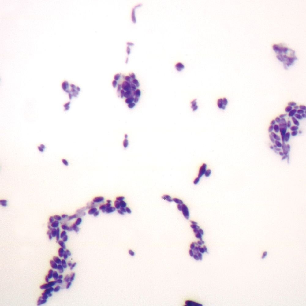
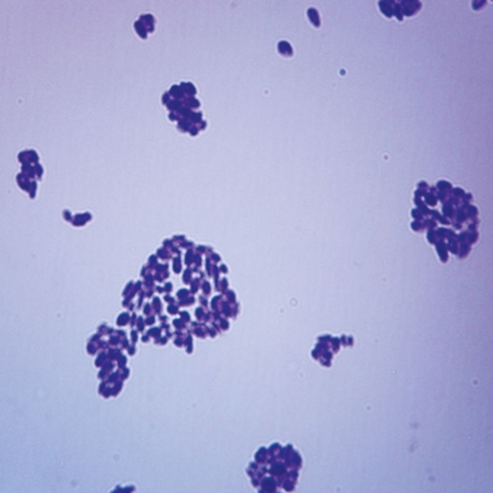
Yeast cells under the microscope appear as single cells or in clusters. They can take on oval or round shapes. Some form long chains called pseudohyphae, especially when in stressful environments.
Depending on their phase of growth, yeast cells might present buds. Buds are a sign of asexual reproduction, known as budding. In this process, a small daughter cell forms on the parent yeast cell.
A clear identification of these cells and their morphologies is invaluable. It lets industries pick the best strains for their products. It also helps researchers understand cellular processes and devise new studies. The keyword ‘yeast under microscope’ encompasses this fascinating exploration into the microscopic world of yeast cells.
Preparing Yeast Samples for Microscopy
To see yeast under a microscope, we need to prep the samples first. This stage is crucial. A good sample gives clear images, revealing the yeast’s detailed structure. Therefore, there are several key steps in this process.
First, we clean the yeast. We need to remove any debris or impurities that might blur the view. Then, we mount the yeast on a slide. We use special stains to make the yeast cells stand out. These stains bond to different parts of the cell. They help us see the shapes and sizes more clearly.
Next, we fix the cells in place. This keeps them from moving or changing while we look at them. We often use heat or chemicals for this part of the process. Finally, we cover the sample with a cover slip. This protects the cells and keeps them flat. Every step is done with care. We want to keep the cells’ details intact during the observation. With the samples prepped right, we’re ready to take a closer look at the yeast under microscope.
Microscopic Techniques for Yeast Observation
Viewing yeast under a microscope requires specific techniques. These approaches expose the tiny details of yeast cells. They vary, from simple light microscopy to advanced electron microscopy.
Light Microscopy allows us to see basic structures. We can observe live yeasts and their movement. This technique is common and cost-effective.
Fluorescence Microscopy uses special dyes. These dyes glow under certain lights. Thus, we can highlight parts of a cell. We see cell walls, nuclei, and other organelles in detail.
Electron Microscopy gives detailed images. It has much higher magnification than light microscopy. This way, we see the yeast’s inner structures. But, it requires a vacuum. We can’t observe living cells with this method.
Confocal Microscopy builds 3D images of yeast. It uses lasers and pinhole cameras. The focus is sharp at a single depth at a time. Layer by layer, we get a full picture.
Using these methods, we explore yeasts in depth. Each technique sheds light on different aspects. Together, they give us a comprehensive view of yeast under a microscope. We understand yeast better and use it more effectively.
Identifying Yeast Cell Structures
To identify yeast cell structures under a microscope, precise observation is key. By focusing on specific characteristics, we can differentiate various components within the cell. Using different microscopic techniques, it’s possible to discern the intricate details. Here’s what we specifically look for when identifying yeast cell structures:
Cell Wall and Membrane
The cell wall is the outermost layer, providing shape and protection. Under a microscope, we spot its robust, clear outline. The membrane inside it, however, is subtle. It encases the cell’s contents and regulates what goes in and out.
Nucleus
The nucleus is the command center. It’s usually a darker, round spot within the cell. Stains often help to make it more visible.
Cytoplasm
Filling most of the cell, the cytoplasm looks like a dense, fluid space under the microscope. It holds various organelles.
Vacuoles
Vacuoles are storage bubbles. In yeast, they’re quite prominent and easily noticed as clear spheres inside the cytoplasm.
Budding Sites
Yeast, known for budding, will often show smaller cells attached to a parent cell. This indicates active cell division.
Mitochondria and Ribosomes
Though harder to see, mitochondria and ribosomes are present as well. Advanced techniques like electron microscopy can reveal these tiny powerhouses and protein builders.
Through identifying these structures, we gain valuable insights into yeast biology. With ‘yeast under microscope’ studies, we can deduce cell health and functionality, aiding both industry and research in their endeavors.
Common Yeast Morphologies
Under the microscope, yeast presents with various shapes, guiding experts in their work. We’ll look at the most common morphologies seen when yeast is observed under a microscope.
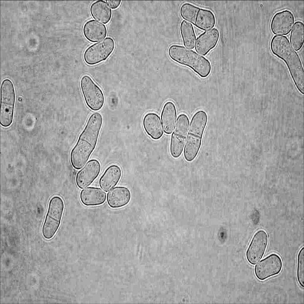
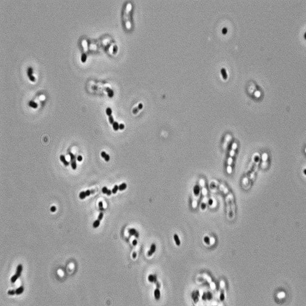
Oval and Round Shapes
The typical shape of Saccharomyces cerevisiae is oval to round. It’s the standard form most people picture when they think of yeast.
Pseudohyphae Formation
Some yeast strains form pseudohyphae, which are chains of elongated cells. This often happens in stressful conditions.
Budding Yeast Cells
A tell-tale sign of yeast reproduction is the budding process. Here, small daughter cells bud off from the parent.
Colonial Morphologya
On solid media, yeast can form colonies with characteristic shapes and textures. This is another aspect of their morphology.
These common shapes and forms are more than just interesting to look at; they’re important clues. They help us understand yeast behavior and its suitability for different uses in industry and research. The term ‘yeast under microscope’ involves exploring these morphologies to gain deeper insights.
Advances in Yeast Microscopy
Yeast microscopy has made great strides in recent years. These advances help us see yeast like never before. New techniques and technology have improved our views into yeast’s world. We now have methods that speed up the analysis and give more detail.
Breakthroughs in digital imaging allow for sharper, higher-resolution images. This means we can observe finer details in yeast cell structures. High-definition cameras and enhanced software play key roles here.
Further, super-resolution microscopy breaks the limits of traditional light microscopy. It allows scientists to see tiny parts of yeast cells not visible before. This technique has pushed our understanding of yeast biology to new levels.
Another advance is live-cell imaging. This lets us watch yeast cells grow and reproduce in real time. We observe their behaviors and interactions closely and constantly.
Automation in microscopy is also a game-changer. It makes it easier to study large numbers of yeast cells. With automated systems, we can collect data faster and with greater accuracy.
Lastly, image analysis software has become more sophisticated. It helps researchers analyze structures and patterns within the yeast cells efficiently. These tools aid in both basic and advanced yeast studies.
In sum, ‘yeast under microscope’ research benefits greatly from these technological leaps. We can now delve deeper into the life of yeast at the microscopic level. The findings from these advanced methods enhance both scientific knowledge and practical applications.
Application of Yeast Microscopy in Industry and Research
The use of ‘yeast under microscope’ extends far beyond simple observation. It has vital applications in industry and research. Let’s take a closer look at how microscopy of yeast shapes these areas.
In the food industry, especially in baking and brewing, observing yeast helps enhance product quality. For example, by studying the fermentation process under the microscope, experts can optimize yeast strains. This leads to better tasting bread and more flavorful beer.
In biofuel production, researchers study yeast to improve the efficiency of fuel generation. This involves analyzing yeast’s ability to convert sugars into ethanol, which is crucial for biofuel.
In the medical field, using a microscope to examine yeast contributes to the understanding of diseases. Since some human cells share similarities with yeast cells, this research can lead to breakthroughs in treatments.
In agriculture, microscopy helps examine the role of yeast in soil health and plant growth. Farmers can use this knowledge to employ natural yeast-based products to enhance crop yields.
Finally, in academic research, observing yeast under a microscope aids in understanding basic cellular processes. This can lead to discoveries about cell biology and genetics that have wider implications.
In short, ‘yeast under microscope’ is a phrase that captures the importance of this tool in advancing numerous sectors. The insights gleaned from microscopy aid in developing new technologies and improving existing processes across various fields.