An Introduction to Ant Anatomy
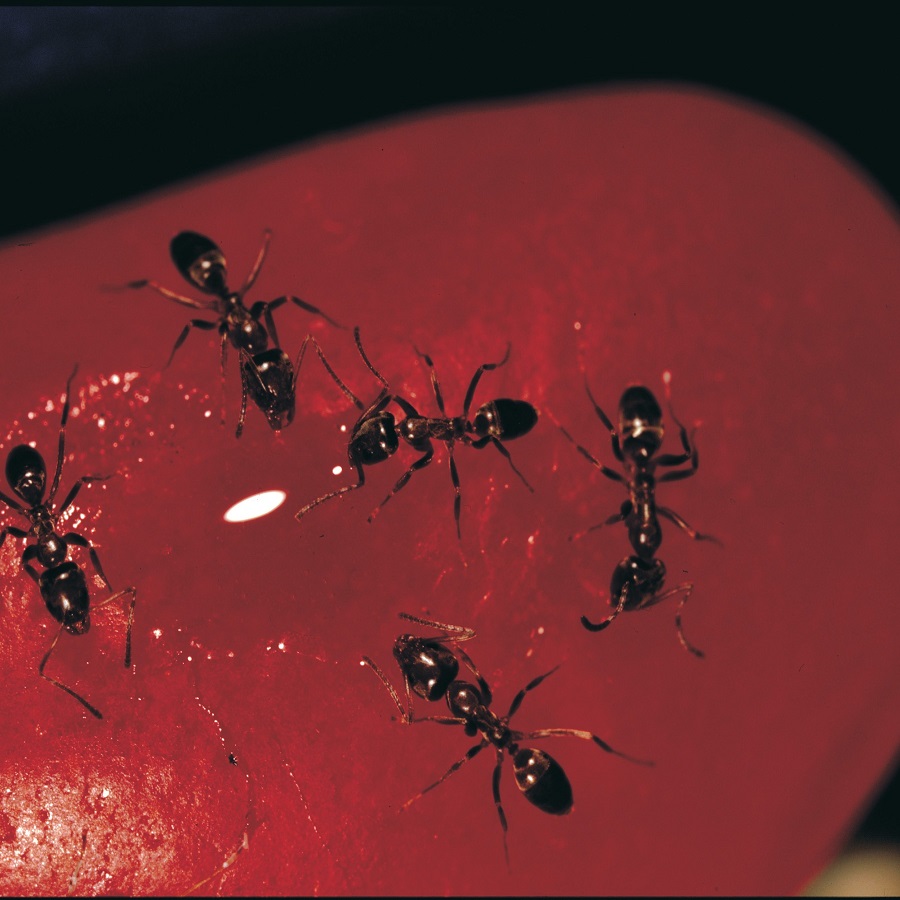
Ants, often seen busily scurrying around, are more than they seem. A closer inspection of these insects presents a fascinating glimpse into a complex anatomical structure. Using a microscope, we can delve into the intricate world of ant anatomy, which supports their diverse roles in ecosystems.
Tiny yet mighty, ants boast a hard exoskeleton. This serves as both armor and support for their bodies. Their three main body parts, the head, thorax, and abdomen, are distinct and perform essential tasks. The head houses critical sensory equipment and impressive mandibles. Meanwhile, the thorax anchors robust legs and, in some species, wings. The abdomen contains vital internal organs.
Examining an ant under a microscope reveals these parts in mesmerizing detail. Microscopes allow us to see the fine hairs, or setae, covering their bodies. These setae have roles in sensation and communication. The jointed appendages, vital for movement and work, also come into focus.
Getting to know ant anatomy is not just academic. It helps us appreciate the adaptability and efficiency of these creatures. In the following sections, we’ll prepare for this microscopic exploration and highlight key features of ant anatomy to observe. Keep your eyes peeled; we’re about to zoom in on an astonishingly small world.
Preparing for Microscopic Examination
Before embarking on the microscopic journey of ant examination, proper preparation is crucial. The following steps will guide you in setting up for a detailed observation of an ant under a microscope.
- Select the Right Microscope: Utilize a stereo microscope, also known as a dissecting microscope, for its lower magnification and larger working area. This is ideal for observing ants.
- Prepare the Specimen: Ideally, opt for a preserved ant specimen. Remember, handling live ants can be challenging and often requires permits.
- Choose Appropriate Magnification: Start with a low magnification to locate your ant on the slide. Then, gradually increase it to observe specific anatomy details.
- Adjust Lighting: Ensure sufficient lighting for clear viewing. Most stereo microscopes come with built-in lighting. You may need additional light sources depending on the model.
- Use Glass Slides and Coverslips: For smaller ants or parts, mount them on a glass slide. Use a coverslip to flatten and secure the specimen.
- Employ Staining Techniques: Some features may need staining to become visible under the microscope. However, this requires extra care, as it can damage the specimen.
- Record Your Observations: Keep a notebook and pen handy to jot down what you see. Document the magnification levels and any peculiarities you notice.
- Clean Up Properly: After you are finished, clean the microscope and tools to keep them in good condition for the next use.
By following these steps, you’ll be well-prepared for an in-depth look at ant anatomy under a microscope and ready to move on to identifying key anatomical features.
Identifying Key Ant Anatomical Features
When you place an ant under a microscope, several key anatomical features become visible. Recognizing these features can enhance your understanding of ant biology and their role in the environment. Here are the primary anatomical parts to look for during your examination:
- Head: The ant’s head is the control center, featuring the eyes, antennae, and mandibles. Under magnification, you can observe the compound eyes, which provide a broad field of vision.
- Antennae: These sensitive appendages on the head allow the ant to sense its surroundings. They are key for communication and navigation.
- Mandibles: Strong and versatile, the mandibles are the ant’s tools for carrying objects, digging, or defense.
- Thorax: The middle part of the ant supports strong legs and, in some species, wings. Examine how the legs attach to the thorax and move.
- Abdomen: The abdomen houses important organs like the digestive system and, in queens, reproductive structures. It’s segmented and often shows distinct texture and coloration under a microscope.
- Setae: Tiny hair-like structures called setae cover the ant’s body. These help in sensing the environment and can be quite prominent depending on the species.
By focusing on these key areas, you’ll gain a deeper appreciation for the complexity of ants. As you adjust the microscope, each feature will provide clues about the ant’s habits and capabilities. Keep your notes close to record unique observations, which may vary from one species to another.
The Exoskeleton and its Role in Ant Physiology
The exoskeleton is a key feature when observing an ant under microscope. This rigid outer shell provides protection against predators and environmental hazards. Not just a shield, it also supports the ant’s body and its movements.
An ant’s exoskeleton is made of chitin, a sturdy material that withstands stress and strain. Under the microscope, you’ll see its segmented nature, which allows for flexibility. This segmentation is essential for an ant’s mobility.
Additionally, the exoskeleton helps prevent water loss. This is vital for an ant’s survival, especially in dry environments. It serves as a barrier, maintaining the internal moisture.
Lastly, the exoskeleton is where muscles attach. When viewing an ant under a microscope, look for the points of attachment. These spots show where the ant’s power to move originates from.
This sturdy structure is integral to ant physiology. It allows them to lift and carry objects many times their own weight. In your microscopic journey, appreciate this marvel of nature that is the ant’s exoskeleton.
Magnifying the Ant’s Head: A Closer Look at the Mandibles and Antennae
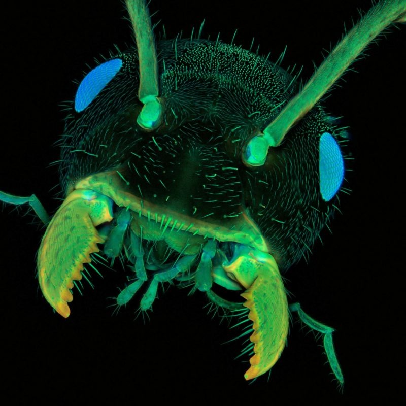
When studying an ant under microscope, the head is particularly intriguing. It’s the command center where all sensory input converges. Through magnification, the head’s intricate details are brought to life, showcasing two critical components: the mandibles and antennae.
Mandibles
The mandibles are the versatile tools of an ant. They are strong and dexterous, allowing ants to cut, carry, and fight. Magnified, you’ll see the sharpness and complexity of their design. This helps us understand how ants can handle such diverse tasks. Look out for the mandible’s edges and points. They reveal how ants can manipulate their environment so efficiently.
Antennae
Next are the antennae. These slender structures sense the world around the ant. Under the microscope, you can view the fine segments and hairs that make up the antennae. They pick up chemical signals, touch, and even sound vibrations. Observe the delicate joints. They give the antennae flexibility to explore surfaces and air.
In our microscopic exploration, the fusion of strength in mandibles and sensitivity in antennae is captivating. It shows us how ants interact with their habitats. Keep note of the size and shape differences among species. These may point to varying habitats and behaviors. Finally, remember our keyword ‘ant under microscope’ is key to this deep dive into the tiny world of ants.
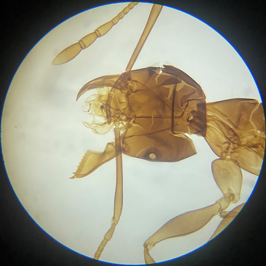
Ant Thorax and Its Appendages
When observing an ant under microscope, the thorax is a notable region. It serves as the connection point for the ant’s vital appendages; its legs and sometimes wings. Let’s take a magnified look at these appendages and their functionality.
Legs
The legs of an ant are a testament to their engineering marvel. They are jointed and robust, allowing for swift, coordinated movement. Through the microscope lens, inspect each leg’s segments: the coxa, trochanter, femur, tibia, and tarsus. Observe how muscles within the thorax anchor to these segments, giving ants their strength to lift or carry. Movements seen under magnification can reveal how each leg functions in unison.
Wings
Only certain ants, like queens and males during mating flights, have wings; worker ants do not. In those ant species, wings connect to the thorax. Under high magnification, you might notice the wing’s thin membrane and the veins that provide structure. These veins appear as delicate patterns that support flight. However, after their nuptial flight, queens shed their wings, making it less common to observe them under a microscope.
The thorax, as seen through a stereo microscope, is a hub of activity for an ant. Note how the thorax’s design supports complex movements and functions. Compare the thoracic structures of different ant species. This can give insight into their lifestyle or living environment. Remember, with each adjustment of the microscope, a new detail of ant anatomy is revealed, deepening our understanding of these fascinating insects.
Exploring the Abdomen: Internal Organs Seen Through the Lens
When viewing an ant under microscope, the abdomen is a focal area, harboring several critical internal organs. Through the lens, you can identify structures like the digestive tract, which processes the ant’s food. A closer inspection might reveal the crop, where food is stored before digestion. Additionally, the heart, a tube-like organ pumping hemolymph (ant’s blood), becomes visible and showcases the circulatory system’s simplicity.
With further magnification, the reproductive organs may come into view, especially in queens. These include the ovaries, where eggs are produced, signifying the ant’s role in colony multiplication. The gaster, the bulbous end of the abdomen, often contains venom glands that contribute to the ant’s defense mechanisms.
Each organ’s condition, size, and relative position provide important biological information. For instance, the size of the venom gland may relate to an ant’s aggression or predatory nature. By detailing these observations and considering each organ’s function, we gain insight into the ant’s survival strategies.
As you turn the focus knob of your microscope, note the abdomen’s segmentation and the flexibility it affords for movement. This examination reveals how the ant’s internal anatomy supports its external actions. It’s a testament to their adaptability in diverse environments. Whether a worker, drone, or queen, understanding the internal structures under the microscope enriches our knowledge of these industrious insects.
Reproductive Organs and the Ant Colony Structure
When you examine an ant under microscope, the reproductive organs take center stage, especially in the queen ants. These organs are crucial for the survival of the colony. Here’s what to look out for when viewing these key features.
Queen Ants’ Reproductive Organs
In queen ants, the reproductive organs are the most significant. Under the microscope, you may be able to see the ovaries. These are where the eggs develop. Fertile queens have larger, more visible ovaries reflecting their vital role in laying eggs. Note the differences in size and structure when comparing queens to workers or males.
Male Ants’ Reproductive Features
Male ants, often called drones, have reproductive parts designed for mating. Their primary role is to fertilize the queen. Magnifying the male ant can uncover the testes or sperm storage areas. However, male ants are often harder to study as they do not live as long after mating.
Colony Structure Insights
The colony’s structure is linked to the reproductive roles of different ants. Workers, which are sterile females, focus on foraging and care for the brood. Studying their anatomy under magnification, you may not find reproductive organs. But, their behavior and physique hint at their roles in the colony. Knowing who has what role helps us understand the ant colony as a whole.
By examining the reproductive organs of ants under a microscope, we learn about the species’ life cycle and society. These insights into the breeding strategies clarify how ant colonies thrive. Each ant, whether queen, drone, or worker, has a place in their complex social structure. So, next time you place an ant under microscope, observe how these minute parts play a massive role in the colony’s success.
Common Ant Species and Microscopic Differences
When performing a close study of an ant under microscope, one quickly realizes the diversity among different ant species. The microscopic examination not only highlights the general anatomy shared by all ants but also the subtle differences that distinguish one species from another. Here, we will overview some common ant species and the unique features visible under magnification that help to identify them.
- Carpenter Ants (Camponotus spp.): Large and robust, carpenter ants often show a distinct, heart-shaped head and powerful mandibles when viewed under a microscope. Their clear segmentation is more noticeable due to their size.
- Fire Ants (Solenopsis spp.): These ants are smaller, with a notorious sting. Under the microscope, look for their two-segmented pedicel, and a stinger visible at their abdomen’s end.
- Pharaoh Ants (Monomorium pharaonis): Pharaoh ants are tiny and light-colored. Microscopic examination reveals their small eyes and a twelve-segmented antenna which is quite long for their body size.
- Pavement Ants (Tetramorium spp.): Distinguished by their grooved head and thorax, pavement ants have a set of spines on the back of their thorax, which can be fascinating to inspect under higher magnification.
- Leafcutter Ants (Atta spp. and Acromyrmex spp.): Renowned for their foliage-ferrying behavior, these ants display large, spiky mandibles under a microscope, helpful for cutting through leaves.
Observing these traits can be enlightening, as different species have adapted physically to their environmental niches and roles within the colony. Microscopic variances like the length of setae, the pattern of veins in the wings, or the shape of the antennae give us clues about their sensory abilities and lifestyle. By learning to spot these nuances, we can identify an ant species and gain a greater understanding of their unique adaptations.