Introduction to Confocal Microscopy
Confocal microscopy stands as a powerful imaging tool in scientific research. Its ability to yield high-resolution, three-dimensional images makes it invaluable in various fields. Unlike traditional microscopy, a confocal microscope utilizes pinpoint laser light to scan samples. This results in sharp images with minimal noise. Researchers rely on this technique to observe fine details within cells and tissues.
At its core, a confocal microscope includes a light source, typically a laser, that illuminates the specimen. A pinhole in an optical path ensures that only light from the focal plane reaches the detector. This setup is crucial for enhancing image clarity and depth. Images obtained from different depths can be reconstructed to create a 3D model of the sample. Such models give scientists insights into the complex structures within biological specimens.
Aided by computer software, a confocal microscope can generate detailed visualizations. These visualizations aid in groundbreaking discoveries across biomedical and material sciences. Researchers can explore the intricate architecture of cells and track dynamic processes in real time. Ensuring that each confocal microscopy session is effective involves understanding the instrument’s mechanics and capabilities. Hence, knowledge of its basic components is essential for any aspiring scientist.

The Basic Components of a Confocal Microscope
Understanding the basic components of a confocal microscope is key to mastering its use. Let’s delve into each of these pivotal elements:
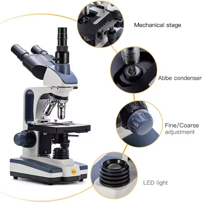
- Laser Light Source: The heart of a confocal microscope is its laser, which provides a focused beam of light. This light is essential in scanning the sample and producing clear images.
- Pinhole Aperture: Located near the detector, the pinhole allows only light from the focal plane to pass. This enhances image resolution and depth perception.
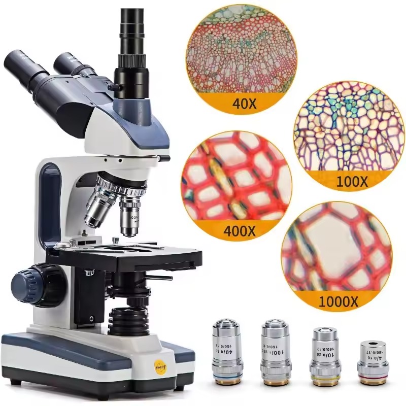
- Objective Lens: This lens collects light from the sample. Its magnification power is crucial for revealing fine details.
- Scanning System: A motorized component that moves the laser beam across the sample. Precise movement is vital for accurate 3D reconstruction.
- Detector: Sensors capture the light passing through the pinhole. They convert it into an electrical signal that creates the image.
- Dichroic Mirror: This mirror reflects specific wavelengths while allowing others to pass. It effectively separates the imaging light from the sample illumination.
- Computer Software: Advanced software processes the signals. It assembles the collected data into a high-resolution image or a 3D model.
Each part of the confocal microscope works in harmony. These elements ensure that researchers can observe specimens with unmatched clarity. In the next sections, we will explore how these components come together to make confocal microscopy work.
How Confocal Microscopy Works
Confocal microscopy operates on a sophisticated yet fundamental principle. A laser emits pinpoint light to illuminate the specimen. The laser scans across the sample systematically, part by part. As it moves, it provides a series of focused light spots. These spots generate the detailed images for which confocal microscopes are renowned.
To capture these images, here’s what happens:
- Focusing the Light: The laser light focuses sharply on a single point in the specimen. This is known as the focal point.
- Eliminating Out-of-focus Light: The pinhole aperture plays a critical role here. It ensures that only light from the focal point reaches the detector. This blocks out-of-focus light and enhances clarity.
- Capturing the Image: The detector captures the light that comes through the pinhole. This light is then turned into an electrical signal to create an image.
- Building the Picture: The scanning system moves the laser spot across the sample. It does so in a raster pattern, similar to how a TV screen is scanned. This creates a two-dimensional image slice by slice.
- Creating 3D Models: By taking multiple 2D images at different depths, a three-dimensional model of the specimen can be created. This is achieved through the z-axis movement control in the microscope.
- Processing the Data: Computer software receives the signals from the detector. It processes these signals to assemble the two-dimensional images into a coherent 3D representation.
Thanks to this process, scientists and researchers can obtain high-resolution images at micron and sub-micron levels. These images are remarkably clear, providing a deeper understanding of the specimen being studied. By mastering confocal microscopy, detailed analysis of cellular structures and their functions is possible. It stands out from traditional microscopy by offering a dynamic view of live processes as they occur in real-time.
Advancements in Confocal Microscopy Technology
Confocal microscopy technology has grown leaps and bounds over recent years. These advancements are critical in pushing the boundaries of what scientists can discover with this tool. Here are several key developments:
- Higher Resolution Imaging: Modern confocal microscope models now feature enhanced optics and digital processing. This results in images with finer detail and better clarity than ever before.
- Faster Scanning Speeds: Improvements in scanning technology have cut down image acquisition times. Faster scans are crucial for observing dynamic processes in living cells.
- Spectral Imaging Capabilities: Newer confocal microscopes can capture images across various wavelengths. This allows for multiplex fluorescent imaging, which is vital for complex biological analysis.
- Advanced Detection Systems: The latest detectors have increased sensitivity. This means they capture low-light samples with greater efficiency, reducing signal noise.
- Multi-Photon Excitation: Some confocal microscopes now employ multi-photon excitement strategies. These techniques enhance penetration depth and reduce phototoxicity in live specimens.
- Automation and Software: We see advancements in user-friendly software and automated systems. They make operating confocal microscopes more intuitive and efficient for research purposes.
The evolution of confocal microscopy technology continues to empower researchers. With these improvements, they achieve unprecedented insight into the microscopic world. As we integrate these enhancements, the scope and accuracy of scientific investigations will continue to expand.
Applications of Confocal Microscopy in Research and Medicine
Confocal microscopy has revolutionized research and medicine through its diverse applications. Here’s how this tool is making an impact:
- Cellular Biology: Researchers study cell structures and functions. They observe how cells interact and behave under different conditions using confocal microscopy.
- Cancer Research: It allows for detailed examination of tumor samples. Scientists analyze cell morphology and monitor tumor progression or response to treatments.
- Neuroscience: Neuroscientists utilize confocal microscopy to probe into the brain. They explore neural pathways and connections, aiding in understanding of brain diseases.
- Developmental Biology: The tool tracks developmental processes in organisms. It reveals how cells and tissues change over time.
- Immunology: Confocal microscopy identifies and locates various immune cells within tissues. This is important for studying immune responses and disorders.
- Clinical Diagnostics: Doctors diagnose diseases by examining tissue biopsies. Confocal microscopy provides high-resolution images necessary for accurate diagnoses.
- Pharmaceutical Research: It evaluates drug effects at the cellular level. Researchers see how drugs interact with cells in real time.
- Material Science: The microscope helps in studying material surfaces and structures. It ensures better understanding of material properties.
Each of these areas benefits from the precise imaging that confocal microscope technology provides. As the technology advances, its applications in research and medicine will likely grow even further. Researchers and medical professionals rely on confocal microscopy to gain valuable insights that drive scientific and medical progress.
Confocal Microscopy vs. Traditional Microscopy
When comparing confocal microscopy to traditional microscopy, several key differences stand out. These distinctions make confocal microscopy a unique and powerful tool in scientific research.
- Focused Laser Scanning: Unlike traditional microscopes that use diffused light, a confocal microscope employs a laser to scan the sample. This provides a focused beam of light for high-resolution images.
- Minimized Noise: The pinhole in confocal microscopy ensures that only light from the focal plane is detected. This technique reduces the noise and enhances the clarity of the images.
- Enhanced Depth: Traditional microscopes give mostly flat images. In contrast, confocal microscopy can capture and stack multiple focal planes. This creates detailed 3D reconstructions of specimens.
- Greater Detail in Live Imaging: Confocal microscopy excels at observing live cells. It allows scientists to monitor cellular processes as they happen. Traditional microscopy does not offer the same level of dynamic imaging.
- Reduced Photodamage: The pinpoint accuracy of the laser means less light exposure for the sample. This is crucial for preserving live specimens, unlike the broader light exposure in traditional microscopy.
- Software Integration: Confocal microscopes are often coupled with advanced computer software. This aids in the complex analysis and 3D modeling of samples, surpassing the capabilities of traditional microscopy.
These advantages explain why confocal microscope use has surged in labs worldwide. Its ability to give clear, detailed views into the microscopic world is unmatched. For cutting-edge research requiring the finest detail and live observation, confocal microscopy is the preferred choice.
Tips for Optimizing Confocal Microscopy in Your Experiments
Maximizing the potential of a confocal microscope in your experiments requires skill and knowledge. Below are practical tips to help you enhance the quality of your confocal microscopy sessions:
- Use the Correct Settings: Begin by adjusting your confocal microscope to the appropriate settings. Match laser power, pinhole size, and detector gain to your sample’s requirements.
- Optimize Sample Preparation: Proper sample preparation is essential. Ensure your specimens are properly fixed and stained for the best imaging results.
- Choose the Right Objective Lens: Select an objective lens with suitable magnification and numerical aperture. This step is crucial for capturing fine details clearly.
- Maintain Clean Optics: Regularly clean the lenses and other optical elements. Dust and fingerprints can greatly affect image quality.
- Manage Laser Power: Too much laser power can damage live samples. Use the minimum power necessary to get a clear image while minimizing photodamage.
- Fine-Tune the Pinhole: The right pinhole diameter enhances resolution. For optimal results, adjust according to the wavelength and your imaging needs.
- Utilize Software Tools: Take advantage of computer software for image processing. Proper use can greatly improve image clarity and detail.
- Prevent Sample Drift: Ensure your sample is securely mounted to prevent movement. Sample drift can lead to blurry images and inaccurate data.
- Optimize Scan Speed: Balance scan speed with image quality. Faster scan speeds can reduce resolution, so find the speed that gives the best clarity without sacrificing time.
- Understand Signal-to-Noise Ratio: Strive for a high signal-to-noise ratio. Adjust detection settings to ensure the clearest possible images.
By implementing these tips, you can significantly improve the performance of your confocal microscopy studies. Always remember to keep learning as confocal microscopy technology advances to stay at the forefront of imaging excellence.
Future Directions in Confocal Microscopy
The future of confocal microscope technology is exciting and promising. As researchers seek even greater insights into the microscopic world, several areas of advancement stand out. Here are some future directions where confocal microscopy may evolve:
- Integration with Artificial Intelligence (AI): AI can manage vast data and refine image analysis. This could lead to faster, more accurate diagnostics and research outcomes.
- Enhanced Resolution and Speed: Ongoing research aims to push the limits of resolution. Speeding up scans without loss of detail is a critical focus. This means clearer images in less time.
- Improved Live Cell Imaging: Techniques to reduce photodamage further are in development. These will enable longer observations of live cells without affecting their viability.
- Expanded Fluorescent Capabilities: The use of new fluorophores and multi-photon techniques will grow. More colors and deeper tissue imaging will become possible.
- Higher Automation: Automating tasks like focus, calibration, and scanning will make confocal microscopes more user-friendly. Labs without specialists could still achieve high-quality results.
The pursuit of these advancements is relentless. Scientists, manufacturers, and software developers are working together. Their goal is to refine and enhance confocal microscopy. With each passing year, the potential for discovery expands, thanks in part to this powerful tool. The confocal microscope has set a high bar in imaging technology. As we look to the future, it is poised to raise that bar even further.