Introduction to TEM Microscopy
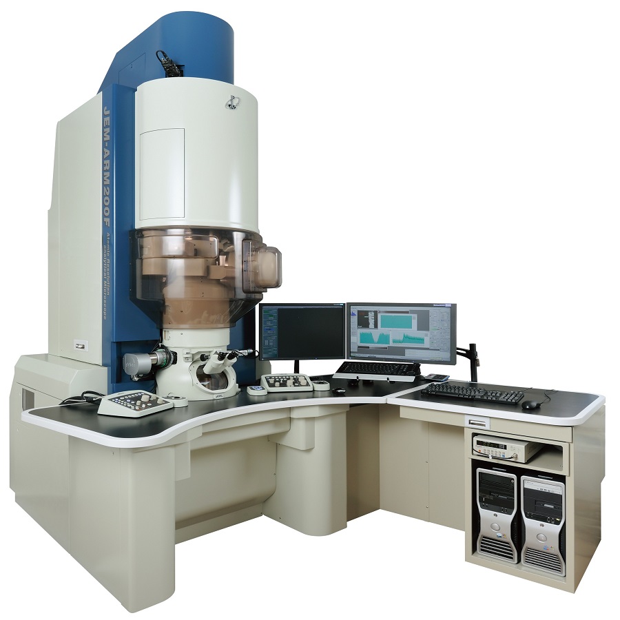
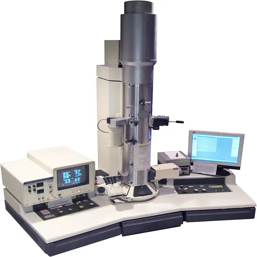
TEM microscopy, or transmission electron microscopy, is a powerful imaging technique. Scientists use it to see at the nanoscale level. The ‘TEM’ in tem microscope stands for ‘transmission electron microscope’. It sends a beam of electrons through a specimen to create an image. This tool has opened up new worlds in scientific research. It reveals tiny details of materials, cells, and other samples.
The tem microscope’s strength lies in its ability to magnify. It surpasses traditional light microscopes in power and clarity. Images from a TEM can show structures as small as atoms. This precision makes it critical for advanced research fields. For example, materials science and biology benefit greatly from TEM.
Users of TEM microscopy must understand its components and physics. Each part of the microscope, from electron gun to detectors, plays a vital role. They work together to produce clear images for detailed analysis. Knowing how the microscope functions can help users capture the best images. It also helps in interpreting the data they provide. We will explore these components and concepts in the following sections.
To sum up, TEM microscopy offers scientists a window into the microscopic world. It allows for groundbreaking discoveries across multiple disciplines. Its capabilities make it an indispensable tool in many areas of research and industry.

Key Components of TEM Microscopes
TEM microscopes rely on several key components. Each plays a critical role in its operation. The electron gun, electromagnetic lenses, and detectors are all crucial. These parts ensure that TEM microscopes produce high-resolution images.
Electron Gun and Its Importance
The electron gun acts as the TEM microscope’s source of electrons. It generates a beam of electrons. These electrons pass through the sample. This is essential for creating the image. Without the electron gun, TEM microscopy would not function.
Electromagnetic Lenses and Image Formation
Electromagnetic lenses focus the electron beam on the sample. They shape the path the electrons take. These lenses magnify the image and help in defining its clarity. They play a part in forming the magnified image we analyze.
Detectors and Image Capture Mechanisms
Detectors in a TEM microscope capture the transmitted electrons. They convert them into a visible image. This image shows the detailed structure of the specimen. The sophistication of the detectors greatly affects image quality.
The Physics Behind TEM
Understanding the physics of TEM microscopy is crucial for mastering its use. At the heart of these complex machines lies the core principles of quantum mechanics that govern the behavior of electrons.
Wave-Particle Duality of Electrons
One fundamental concept in TEM is the wave-particle duality of electrons. This duality refers to the fact that electrons exhibit properties of both waves and particles. In the context of TEM, this means electrons can be focused like light waves using electromagnetic lenses. But, they also interact with the sample like particles. The wave aspect allows the tem microscope to capture high-resolution images by overcoming the limitations of traditional optics.
Electron Scattering and Resolution Limits
The resolution of a TEM is impacted by electron scattering. As the electron beam interacts with the sample, electrons scatter in different directions. This scattering is influenced by the thickness and atomic structure of the specimen. It limits how closely the beam can focus and ultimately defines the resolution of the image. TEM microscopes face resolution limits due to these scattering effects, but advancements in technology continue to push these boundaries. This is essential for scientists needing to visualize samples at the atomic level.
Applications of TEM Microscopy
TEM microscopy has become an indispensable tool across various fields.
Materials Science and Nanotechnology Research
In materials science, TEM microscopes are vital. They allow researchers to observe the structure and properties of materials at the atomic level. This is crucial for developing new materials with improved performance. In nanotechnology, TEM helps in the study of nanoparticles and nanostructures. The tem microscope’s precision aids in designing devices and materials with nanoscale features.
Biological Specimen Imaging
Biologists use TEM to study cells, viruses, and proteins in great detail. It enables the visualization of structures within biological specimens that light microscopes cannot see. This deep look into the cellular level helps in understanding diseases and developing treatments.
Industrial and Semiconductor Analysis
TEM is also important in the industrial sector, especially in semiconductor manufacturing. The technique is used for analyzing defects and for quality control of microchips. Engineers rely on the tem microscope to refine processes and enhance the performance of electronic components.

Advancements in TEM Technology
TEM microscopy has evolved significantly since its inception. Each advancement brings more detailed insights into the nanoscopic world.
Improved Resolution with Aberration Correction
One major stride in tem microscope capabilities is aberration correction. This technique corrects lens imperfections that blur images. Now, TEM users enjoy sharper, more accurate images. Improved resolution means seeing atomic details with unprecedented clarity. This advancement has a big impact on materials science and nanotechnology. It pushes the TEM limits, allowing deeper exploration into the structure of materials.
Cryo-TEM Innovations and Biological Insights
Cryo-TEM is another groundbreaking innovation. This technique freezes samples rapidly, preserving them in a natural state. This is essential for biological specimens which can be sensitive to traditional TEM preparation. With cryo-TEM, researchers observe life’s building blocks without distortion. This has led to incredible discoveries in cell biology and biochemistry. Understanding viruses and proteins at a molecular level has never been clearer. Cryo-TEM offers a window into living systems that was once impossible to achieve.
Comparison Between TEM and Other Microscopy Techniques
Comparing TEM with other microscopy methods illuminates its unique benefits. Scientists often weigh the capabilities of different tools for their research needs. Besides TEM, scanning electron microscopy (SEM) and optical microscopy are popular choices.
SEM vs TEM: Understanding the Differences
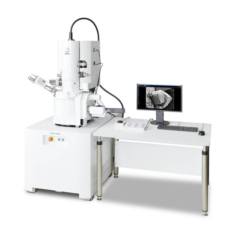
SEM scans the surface of a sample with electrons, revealing topographical details. TEM, on the other hand, transmits electrons through a specimen, showing internal structure. TEM can image at a higher resolution compared to SEM. This is because TEM examines thin sections, revealing atomic details. SEM gives a good 3D surface view but does not offer the same depth of information.
Optical Microscopy and Its Limitations Compared to TEM
Optical microscopy uses light to magnify images and is well-suited for general purposes. However, its resolution is limited by the wavelength of light. This limitation means that optical microscopes cannot view atoms or molecules. TEM surpasses these limits. It uses electron wavelengths much smaller than light. This is why TEM can visualize features down to the atomic level. Optical microscopy remains a tool for larger, visible structures, while TEM excels at the nanoscale.

Maintenance and Operational Challenges of TEM
Like all sophisticated equipment, TEM microscopes require regular maintenance to function optimally. Users must tackle various operational challenges to maintain image quality and prolong the microscope’s lifespan.
Routine Cleaning and Calibration
Regular cleaning of the TEM microscope is crucial to prevent buildup that can interfere with performance. Calibration ensures that the microscope’s components, like electromagnetic lenses, are finely tuned for accurate imaging. Cleaning should be gentle and precise, following manufacturer guidelines. Calibration routines may involve adjusting lens currents and correcting alignments.
Addressing Common Technical Issues
Technical issues in TEM can range from electron beam instability to vacuum system problems. Users need to identify and fix these issues, often with the help of specialized technicians. Regular checks of the filament, alignments, and software are necessary. Swift action is key when technical faults arise to avoid damage and downtime.
Conclusion: The Future of TEM Microscopy and Its Impact on Science
TEM microscopy stands at the forefront of scientific discovery. Its future seems just as promising as its past. Advances in tem microscope technology will likely push research forward in leaps.
We expect to see even higher resolution and more user-friendly TEMs. This could help to unravel complex materials and biological systems. Improved software might make TEM microscopy more accessible to a broader range of scientists. It could simplify image analysis, too.
Innovations in sample preparation will transform TEM’s impact on biology. Methods that are gentler and preserve samples better are on the horizon. New materials might also improve the efficiency of electron detection, sharpening images.
The tem microscope will keep proving itself as key in industries like semiconductors. As devices get smaller, TEM will be crucial for quality control and troubleshooting. In the realm of materials science, expect the unexpected. Insights from TEM will likely lead to new materials with game-changing properties.
In education, TEM may become a more prevalent teaching tool. With tech advances, it could be easier for students to explore the nanoworld.
Finally, the convergence of TEM with other scientific methods could open new research avenues. Scientists might combine TEM with computer modeling or other imaging techniques. This would give even clearer insights into the microscopic world.
To sum up, the tem microscope will continue to shape the future of science in profound ways. Its ongoing evolution ensures that it will remain indispensable in our quest to understand and innovate at the tiniest scales.