The Basics of DNA Structure
Before delving into the various techniques for DNA visualization, it’s crucial to have a fundamental understanding of DNA structure. DNA, or deoxyribonucleic acid, is the hereditary material in humans and almost all other organisms. Every cell in a person’s body contains DNA, which carries the instructions needed for the development, functioning, growth, and reproduction of all life forms.
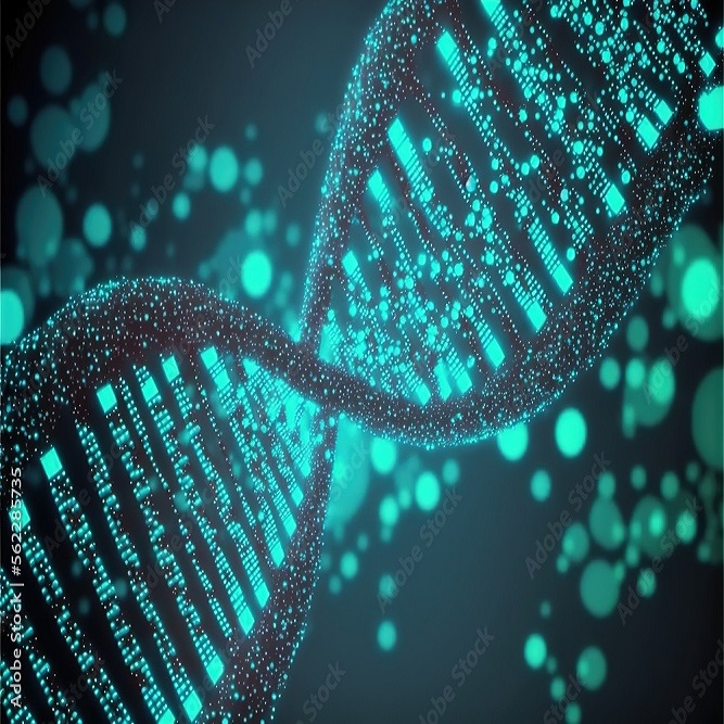
The structure of DNA is often described as a double helix, a term coined by James Watson and Francis Crick who discovered its shape in 1953. Picture a twisted ladder, with the ladder’s steps made of pairs of nitrogenous bases and the sides representing sugar-phosphate backbones. These bases pair in a specific way: adenine (A) with thymine (T), and cytosine (C) with guanine (G).
The sequence of these bases along the DNA strand encodes genetic information. Using dna under microscope techniques like fluorescent staining and electron microscopy, scientists can visualize and study this genetic material. This has revolutionized our ability to understand and manipulate genetic information, leading to breakthroughs in medicine, forensics, and more. But before these advanced viewing methods can be applied, proper preparation of DNA samples is necessary, an aspect we’ll explore in the following sections.
Techniques for DNA Visualization
Visualizing DNA with the right technique can unveil detailed insights into its structure and function. Advanced methods have been developed to observe DNA at the molecular level.
Fluorescent Staining
Fluorescent staining is a popular method for highlighting DNA under a microscope. It involves using dyes that attach to specific parts of the DNA molecule. These dyes shine brightly when exposed to certain wavelengths of light. This illumination allows researchers to see the twists and turns of DNA strands in vivid colors. Fluorescent staining is key in various genetic analyses, helping to identify genetic disorders and map out chromosomes.
Electron Microscopy
Electron microscopy offers a closer look at DNA. This technique uses a beam of electrons instead of light to create an image. The high resolution of electron microscopy makes it possible to examine the structure of DNA in fine detail. Scientists can pinpoint where molecules connect and how they interact with one another at the atomic level. It is crucial for understanding DNA’s physical properties and how these relate to its biological functions.
X-ray Crystallography
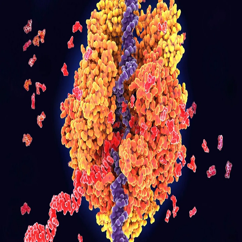
X-ray crystallography provides a different angle on DNA visualization. It involves crystallizing the DNA and then bombarding it with X-rays. The way the rays diffract off the crystal lattice allows scientists to deduce the structure of DNA. This technique was instrumental in discovering the double helix shape of DNA. Today, it remains essential for studying the molecular structure and for the development of new drugs.
Each of these techniques, from staining to bombarding with X-rays, adds to our understanding of DNA. The choice of method depends on the specific details researchers wish to uncover.
The Process of Preparing DNA Samples for Microscope Analysis
Before DNA can be scrutinized under a microscope with techniques like fluorescent staining, electron microscopy, or X-ray crystallography, it must first undergo precise preparation. Here’s a look at the typical steps involved.
- Extraction: The first step involves extracting DNA from cells. This is usually done by breaking open the cell membrane and nuclear envelope using detergents or enzymes.
- Purification: Following extraction, DNA requires purification to remove proteins, lipids, and other cellular debris. Ethanol precipitation is a commonly used method for this purpose.
- Concentration: Once purified, the DNA solution may be too dilute for analysis. Therefore, concentration methods like evaporation or centrifugation are employed.
- Quantification: Before visualization, it’s vital to determine the DNA concentration to ensure clarity. Spectrophotometry is a favored technique for quantification.
- Staining: If using fluorescent staining, appropriate dyes are added to the DNA. These dyes are what make DNA visible under specific wavelengths of light during microscopy.
- Sample Mounting: Finally, the stained or treated DNA is carefully mounted on a slide or specimen grid, ready for examination under the microscope.
Each step must be carried out with care to preserve DNA integrity and ensure accurate visualization. Failure in even one step can lead to poor DNA visibility under the microscope, impacting subsequent analysis.
Advances in DNA Imaging Technology
Advancements in DNA imaging technology have greatly enhanced our ability to study genetic material. These innovations enable researchers to observe DNA under a microscope with unprecedented detail and accuracy. Here are some of the latest advancements that have had a significant impact:
- Super-Resolution Microscopy: This technique surpasses the limits of traditional light microscopy. It allows scientists to view structures as small as a few nanometers, such as DNA strands.
- Atomic Force Microscopy (AFM): AFM can image DNA molecules without staining them. It uses a probe to feel the surface and create images at the atomic level. This method shows the DNA’s shape and volume.
- Cryo-Electron Microscopy (Cryo-EM): Cryo-EM freezes DNA samples at very low temperatures. This preserves their natural shape and allows for high-resolution imaging of DNA in its native state.
- Live-Cell Imaging: Scientists can now watch DNA interact inside living cells in real time. This is crucial for understanding how DNA functions in life processes.
- Fluorescence in situ Hybridization (FISH): FISH attaches fluorescent probes to specific DNA sequences. It helps identify the location of genes within the DNA strand.
- Third-Generation Sequencing Technologies: These offer not just DNA visualization but also rapid sequencing. They help in detecting DNA changes at the single-molecule level.
These advances make it easier to understand the complexities of DNA. They guide us in fields like genetics, molecular biology, and disease research. As these technologies evolve, they promise to reveal even more details about the intricate world of DNA.
Applications of DNA Visualization in Research
DNA visualization plays a pivotal role in expanding our understanding of genetics and molecular biology. There are numerous applications that benefit from the advancements in DNA imaging technologies. Here’s a brief look into how viewing DNA under a microscope is employed in various research arenas.
- Genetic Disorder Diagnosis: Detecting abnormalities in DNA structure facilitates the diagnosis of genetic disorders. By using fluorescent staining and other imaging methods, researchers can identify mutations that cause diseases.
- Drug Development: Insights into the DNA’s molecular structure, often gained through techniques like X-ray crystallography, are vital for designing new drugs. Visualization aids in understanding how drugs interact with DNA.
- Cancer Research: DNA visualization helps in identifying the genetic changes associated with cancer. This knowledge is crucial for developing targeted therapies and personalized treatment plans.
- Forensic Science: Forensic experts use DNA visualization methods to analyze biological evidence. This can assist in criminal investigations and legal matters by comparing genetic material from crime scenes with suspects.
- Evolutionary Biology: By studying the DNA from different species, scientists can track evolutionary relationships. Microscopic visualization techniques help in outlining genealogical ties.
- Agriculture: DNA imaging is used to genetically modify crops for better yield and disease resistance. Understanding DNA structure under a microscope allows for manipulation that supports sustainable agriculture practices.
Each of these applications shows the critical importance of DNA visualization. Advancements in microscopy and imaging technologies have made these applications more precise and effective, providing a clearer picture of DNA’s role in health, development, and the environment.
Ethical Considerations in DNA Imaging
When we look at DNA under a microscope, ethical questions arise. These concern privacy, consent, and the use of genetic data. Let’s consider these critical issues.
Privacy Concerns
Peering into the genetic code raises privacy risks. Individuals may fear misuse of their genetic information. This includes discrimination in jobs or insurance. Safeguarding personal data is vital. Clear guidelines on who can access and use DNA data are necessary.
Informed Consent
Before obtaining DNA samples, informed consent is crucial. People must understand the purpose and potential risks. They should know how their genetic information will be used. Ensuring voluntary participation is also important for trust.
Genetic Data Handling
Handling and storing genetic data is a major concern. It raises questions about who owns this data. Proper handling procedures prevent unauthorized access and misuse. Strict protocols and secure databases are key for protection.
Ethical Research Practices
Research involving DNA must follow ethical guidelines. This includes respecting participant rights and seeking ethical review. The benefits of research should outweigh any risks to individuals.
DNA and Law Enforcement
Use of DNA in forensic science is complex. It helps solve crimes but also involves ethical dilemmas. Maintaining the right balance between solving crimes and protecting privacy is a challenge.
In summary, DNA imaging technology offers great benefits. However, it is essential to consider ethical questions. Respecting privacy, obtaining consent, secure data handling, and ethical research practices are at the forefront. Balancing these concerns is key to using DNA imaging responsibly.
Future of DNA Visualization Technologies
As we peer into the future of DNA visualization, we anticipate breakthroughs that will push the boundaries of what’s possible. Scientists and technologists are working tirelessly to enhance our ability to view DNA under a microscope with even greater clarity and precision. The advent of new technologies will likely provide insights that are not just deeper, but also more accessible and rapid.
Next-Generation Microscopy Techniques
Investigators continue to innovate microscopy techniques for sharper, more detailed DNA images. Techniques that surpass the current capabilities of super-resolution microscopy, providing even more granular detail, are on the horizon. This may lead to observing DNA interactions in real-time, without the need for sample preparation that could alter its native state.
Enhanced Staining Methods
Researchers are developing novel fluorescent staining methods. These aim for higher specificity, allowing clear distinction between different DNA regions. Enhanced staining would improve the accuracy of detecting and analyzing genetic mutations.
Improved Imaging Software
Advancements in imaging software will play a vital role. Software enhancements will facilitate better analysis and interpretation of DNA visualization results. They will make complex data easier to understand and share among researchers.
Accessibility and Portability
Efforts also gear towards making DNA visualization tools more accessible and portable. This could democratize DNA analysis, enabling it to perform in remote areas or in smaller labs without the need for large, expensive equipment.
Integration with Artificial Intelligence
Artificial intelligence (AI) is set to transform DNA visualization, with algorithms that can quickly and accurately analyze DNA structures. AI could predict how mutations affect genetic functions, aiding in the rapid diagnosis and treatment of genetic disorders.
The future of DNA visualization is promising. It will likely enhance our fundamental understanding of genetics, providing new ways to fight diseases, improve forensic analysis, and advance scientific discoveries. The integration of new technologies in DNA microscopy promises exciting developments in the years to come.