Introduction to Smooth Muscle Tissue
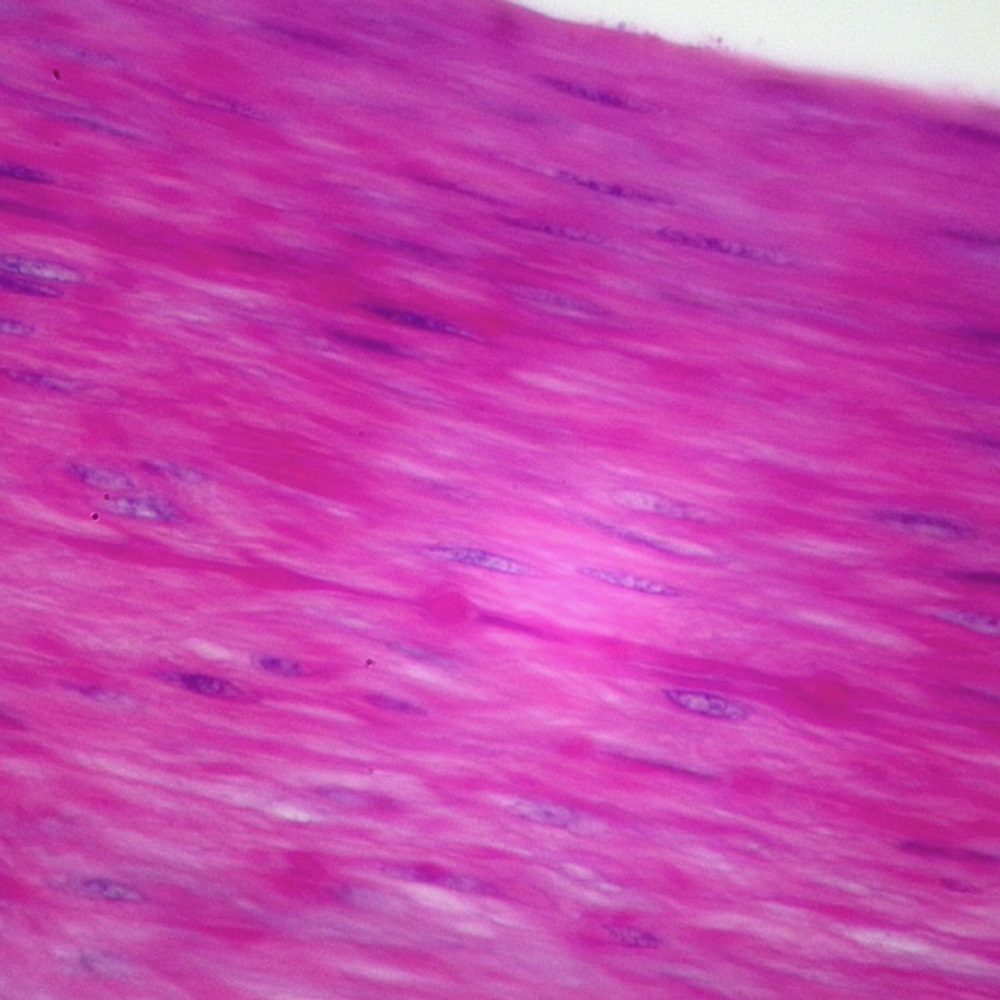
Smooth muscle tissue is a type of muscle found in various parts of the body. Unlike the striated pattern of skeletal muscle, smooth muscle has a uniform, non-striated appearance. It lines the walls of blood vessels and organs, such as the stomach, intestines, bladder, and uterus. This muscle tissue is involuntary, meaning it operates without conscious control. When looking at smooth muscle under a microscope, one can observe its elongated, spindle-like cells. Each cell contains a single nucleus located in the center and is surrounded by a fine network of connective tissue. In this visual guide, we will explore the intricate details of smooth muscle cells and their organization when magnified. This examination will help us understand their vital role in the functioning of various organ systems.
Key Characteristics of Smooth Muscle Cells
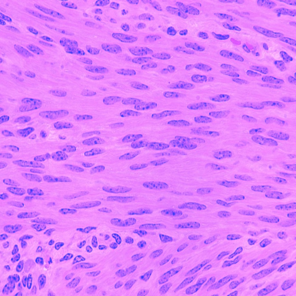
Smooth muscle cells are the building blocks of smooth muscle tissue. These cells display several key features that distinguish them from skeletal and cardiac muscle cells. First, smooth muscle cells have a spindle-shaped, elongated form. This unique shape is very different from the cylindrical and branched forms of skeletal and cardiac muscle cells, respectively.
Another defining characteristic is the single, centrally located nucleus each smooth muscle cell contains. In contrast, skeletal muscle cells can be multinucleated, and cardiac muscle cells usually have one nucleus that is more centrally located.
Smooth muscle cells also exhibit fewer contractile fibers compared to their skeletal and cardiac counterparts. These fibers, which include actin and myosin, are essential for the contractile ability of the muscle but are not organized into the sarcomeres found in striated muscle. This gives smooth muscle its non-striated, homogeneous appearance when viewed under a microscope.
Lastly, the presence of a supportive framework called the extracellular matrix is notable. This matrix surrounds the smooth muscle cells and provides a conduit for signaling and nutrients, which is vital for the cells’ function and maintenance.
By examining smooth muscle under a microscope, researchers can observe these characteristics in detail, aiding in their understanding of how smooth muscle functions within the body. Whether it’s controlling the diameter of blood vessels or facilitating the movement of food through the digestive system, the unique properties of smooth muscle cells are key to their role.
Microscopic Structure of Smooth Muscle
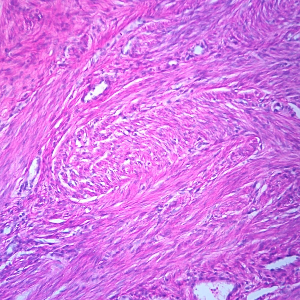
When delving into the microscopic structure of smooth muscle, one can appreciate its complex yet systematic organization. Under the microscope, smooth muscle cells present a tightly packed arrangement. This closeness is necessary for the tissue to exert force uniformally and effectively across the affected organ.
Each smooth muscle cell connects to its neighbors via a type of junction known as gap junctions. These junctions facilitate the swift and coordinated transmission of electrical signals. This is crucial for their synchronous contractions, especially during processes like peristalsis in the digestive system.
Unlike striated muscles, smooth muscle does not have visible banding patterns under the microscope. This lack of striation is due to the even dispersion of contractile proteins throughout each cell. Actin and myosin filaments crisscross at various angles, a setup that is often referred to as a ‘lattice’ structure within the scientific community.
Additionally, smooth muscle cells contain dense bodies, analogous to Z-discs in striated muscle. These dense bodies anchor the actin filaments, allowing for contraction even when these cells stretch considerably. Researchers examining smooth muscle under microscope notice that, during contraction, the dense bodies and lattice structure of filaments enable the cell to contract in a corkscrew-like fashion. This permits the fine-tuned control of tension and length necessary for the varied functions of smooth muscle organs.
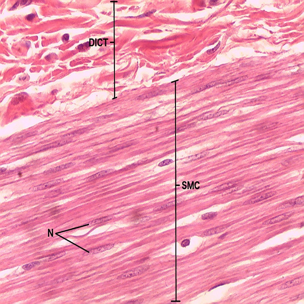
In the intercellular space, there is abundant connective tissue known as the endomysium. This tissue not only supports and binds the smooth muscle cells together, it also holds important blood vessels and nerves. These structures are vital for delivering oxygen, nutrients, and neurotransmitters to the muscle tissue. Under the microscope, this supportive network is visible as a delicate web enfolding the muscle fibers.
Observing smooth muscle under a microscope reveals a highly adapted structural organization, key to its role in involuntary and rhythmic movements throughout the body. Given the right magnification, the intricate interplay of cells and extracellular components affirms the unique capabilities of this muscle type.
Examining Smooth Muscle Under the Microscope
To truly appreciate the complexity and function of smooth muscle, we must look at smooth muscle under a microscope. This magnified perspective offers a detailed view of the cells’ features and how they work together. During microscopic examination, scientists can witness the interaction between the smooth muscle cells and their environment.
When examining smooth muscle under the microscope, we notice several important aspects. The cells appear closely packed, which is vital for their collective force application. Each cell connects to its neighbors through gap junctions, ensuring synchronized contractions. This is especially important for processes such as food movement in the gut.
The lack of striation stands out when viewing smooth muscle tissue. This is because the contractile proteins, actin and myosin, spread evenly in each cell. These filaments intersect at different angles, creating a lattice-like arrangement crucial for muscle flexibility and contraction.
Under high magnification, one can see dense bodies within the cells. These serve as anchor points for actin and contribute to the muscle’s ability to contract in multiple directions. Researchers look for this feature to understand how smooth muscle can maintain tension. It also explains their expansive function in the human body.
Finally, the extracellular matrix appears as a fine network surrounding the cells. It secures the muscle fibers, hosting vital blood vessels and nerves. These structures deliver essential nutrients and signals for muscle function. This intricate network is critical for the survival and performance of smooth muscle tissue.
Microscopic examination of smooth muscle reveals its unique structure and operational mechanism. With each magnified view, the expertise of cell organization and the muscle’s adaptability in various physiological roles becomes clearer. This examination is key in understanding not just the structure but also the very nature of smooth muscle tissue and its indispensable actions in the body.
Differences Between Smooth, Skeletal, and Cardiac Muscle Tissues
When we look at smooth muscle under a microscope, we see a stark contrast to both skeletal and cardiac muscle tissues. Each of these muscle types plays a unique role in the body. Here, we’ll compare their major differences in structure, control, and function.
Smooth muscle is not striated, unlike skeletal and cardiac muscles which show visible banding patterns. This non-striated property of smooth muscle is due to the even spread of contractile proteins throughout the cell. This allows for a range of motions and contractions not typical in the other two muscle types.
Control wise, smooth muscle operates involuntarily. This means it doesn’t require conscious effort to function. In contrast, skeletal muscle is under voluntary control, so we can move it at will. Cardiac muscle, found only in the heart, is also involuntary but it has its own built-in rhythm.
Lastly, the function of these muscles varies greatly. Smooth muscle is found in the walls of hollow organs and helps move substances through the body, like food in the digestive tract. Skeletal muscle enables movement of bones and the body overall. Cardiac muscle works tirelessly to pump blood throughout the body.
In summary, the main differences lie in their appearance under the microscope, control mechanism, and their specific roles in the human body.
Role and Function of Smooth Muscle in the Human Body
Smooth muscle plays a critical role in our body’s daily functions. Found within the walls of hollow organs, it enables the movement and control of various substances. Let’s delve into the primary roles and functions of smooth muscle in different body systems.
Digestive System
In the digestive tract, smooth muscle is crucial for peristalsis. This is the wave-like motion that moves food along for digestion. Without smooth muscle, essential digestion and nutrient absorption would stall.
Circulatory System
Smooth muscle in blood vessel walls helps regulate blood pressure and flow. By contracting and relaxing, it adjusts the diameter of the vessels, controlling the circulation of blood throughout the body.
Respiratory System
The airways in our lungs rely on smooth muscle. It adjusts the airflow during breathing, contracting or relaxing to let more or less air through.
Urinary System
In the bladder, smooth muscle enables the storage and expulsion of urine. It stretches to hold urine and contracts when it’s time to urinate, ensuring proper elimination of waste fluids.
Reproductive System
In females, smooth muscle is integral to the uterus, especially during childbirth. It contracts rhythmically to aid delivery. It also plays a role in controlling the flow of menstrual blood.
Overall, smooth muscle’s ability to contract and relax without our conscious control makes it indispensable for maintaining many critical bodily functions. Its operations are essential for the health and well-being of several major organ systems.
Common Diseases and Disorders Affecting Smooth Muscle
Smooth muscle is crucial for many body functions, but it can be prone to certain diseases and disorders.
Asthma
Asthma affects the respiratory system’s smooth muscles, making it hard to breathe. Muscles around the airways tighten and cause wheezing.
Irritable Bowel Syndrome (IBS)
IBS impacts the digestive tract’s smooth muscles, leading to pain, bloating, and irregular bowel movements.
Hypertension
In hypertension, blood vessel smooth muscles face constant high pressure. This can lead to serious heart problems over time.
Organ Prolapse
Organ prolapse, often in the pelvic region, involves weakened smooth muscles. Organs like the bladder or uterus can slip from their places.
Achalasia
Achalasia is a rare disorder where the esophagus’s smooth muscles fail to relax, making swallowing difficult.
These conditions highlight the vital nature of smooth muscle in bodily functions. When disrupted, they can lead to discomfort and more severe health issues. It’s important to understand these disorders as they affect various body systems regulated by smooth muscle.
Advancements in Microscopic Imaging of Smooth Muscle Tissue
The study of smooth muscle under microscope has seen remarkable advancements. These developments have improved our understanding of its form and function. High-resolution imaging techniques now allow us to observe smooth muscle at the molecular level. This detail provides clues to how these muscles work. Techniques like fluorescence microscopy light up proteins within cells. This helps us see how they move and interact. Electron microscopes offer even more detail, showing the texture of the muscle. We now know how smooth muscle changes during disease. Live-cell imaging techniques track smooth muscles in real time. This shows us how they react to stimuli. New imaging tech also aids in the study of drug effects on smooth muscle. Scientists can now see how treatments change muscle behavior. These breakthroughs in microscopy support research into smooth muscle disorders. Deep understanding of these muscles can lead to better treatments. Advances in imaging contribute to progress in medical science. They help us grasp the complex nature of smooth muscle tissue.