Introduction to Sperm Microscopy
When we talk about looking at sperm under a microscope, it’s a window into the complex world of human reproduction. Sperm play a critical role in procreation, and their study can reveal much about human health and fertility. Sperm microscopy is the detailed examination of spermatozoa using a microscope to gain insight into their structure and function.
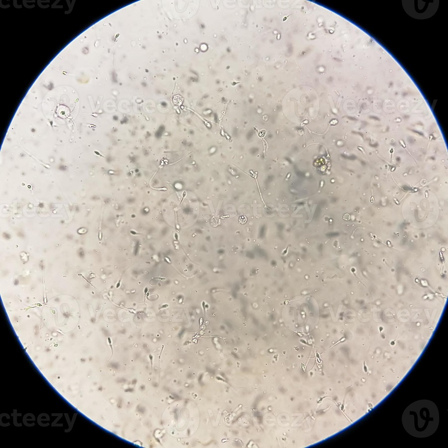
The process involves preparing a sperm sample, placing it under the microscope, and observing the cells. What you see can vary widely. Some sperm may swim quickly across the field of view. Others might drift or be immobile. Scientists and fertility experts use sperm microscopes to evaluate sperm health and diagnose reproductive issues.
Microscopy allows for the assessment of sperm motility, which is a key factor in fertility. It also lets experts inspect the morphology, or shape, of sperm cells. These observations help identify potential fertility problems or diseases. Furthermore, it is a critical step in understanding the outcome of various medical treatments on reproductive health.
For anyone interested in the science of fertility or in professions related to reproductive health, understanding the basics of how we look at sperm under a microscope is fundamental. This knowledge aids in the evaluation of male fertility, the research into reproductive disorders, and contributes to advancements in medical sciences related to human conception.
In this blog, we will journey through the fascinating details of sperm under the microscope, from their anatomy and movement to the techniques and ethical considerations of studying them. Let’s take a deeper look into the hidden world of sperm viewed through a microscope.
The Anatomy of Sperm Observed Under the Microscope
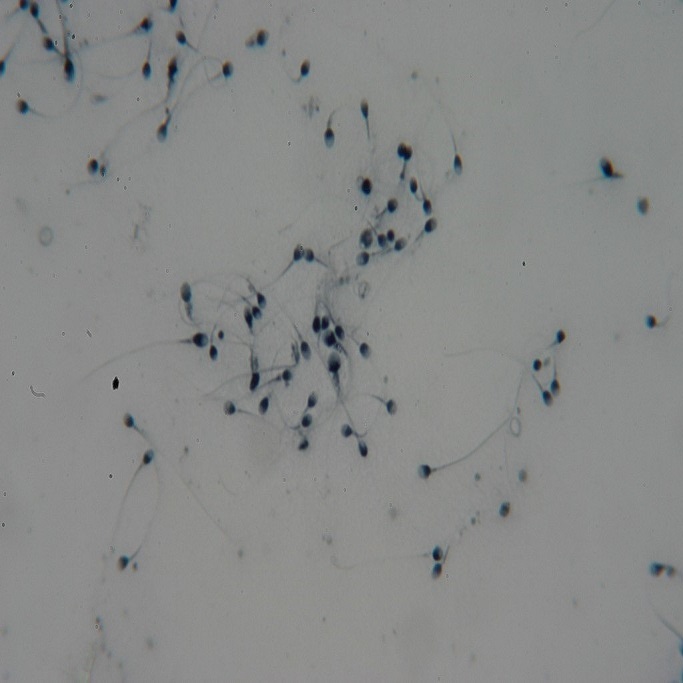
When we observe sperm under a microscope, its complex anatomy becomes apparent. Each spermatozoon, the scientific term for a sperm cell, consists of three main parts: the head, the midpiece, and the tail.
The Head
The head of the sperm is almost oval-shaped and it carries the genetic material. It contains the nucleus, which holds the DNA, and the acrosome, a cap-like structure. The acrosome contains enzymes that help the sperm penetrate the egg during fertilization.
The Midpiece
Beneath the head is the midpiece, which has a central role in powering the sperm cell. It is packed with mitochondria, the cell’s powerhouses. These tiny structures provide the energy needed for the sperm to swim towards the egg.
The Tail
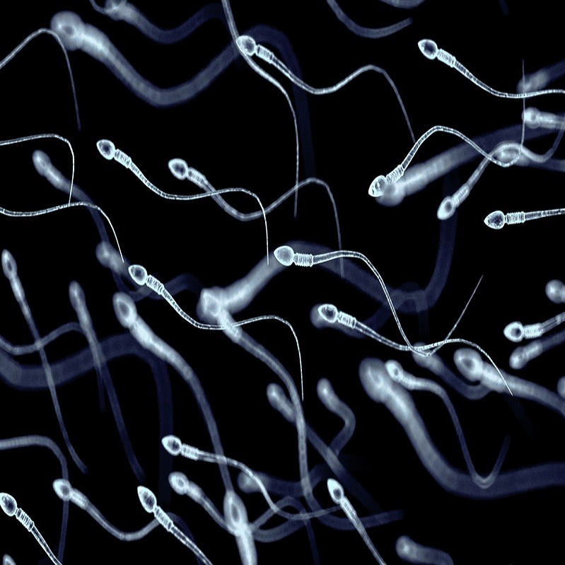
The tail of the sperm, also known as the flagellum, is long and whip-like. It executes the swimming movements that propel the sperm forward. This is a key part of sperm motility, essential for reaching the egg. Thanks to the tail, sperm can move in a straight line or in a corkscrew-like pattern.
Observing the anatomy of sperm under a microscope allows us to better understand how these cells are designed for their ultimate purpose of reaching and fertilizing an egg. The way each part functions is critical to the sperm’s success in reproduction.
Preparing a Sperm Sample for Microscopy
Before viewing sperm under a microscope, proper preparation of the sperm sample is necessary. This ensures clear, accurate observations. Here are the steps typically followed to prepare a sperm sample for microscopy.
- Collection: The first step is collecting the sperm, often through masturbation. The sample should be captured directly into a sterile container to avoid contamination.
- Liquefaction: After collection, the sperm sample undergoes liquefaction. This process usually takes about 15 to 30 minutes at room temperature. It allows the semen to transform from a gel-like state to a liquid. This change helps in analyzing the sperm under a microscope.
- Concentration and Washing: Next, scientists may concentrate the sperm by spinning the sample in a centrifuge. The sperm settles at the bottom, forming a pellet. The technicians then wash the sperm to remove contaminants and chemicals.
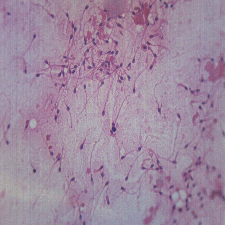
- Staining: Sometimes, a dye or stain is added to the sperm to make certain structures more visible. This is particularly useful for observing the head and tail.
- Smear and Fixation: A small amount of the washed sperm is spread on a microscope slide to create a thin smear. The sperm sample is then fixed on the slide. Fixation locks the sperm into place and preserves their structure.
- Drying: After fixation, the sperm smear is air-dried. The dry sample ensures clarity and prevents the sperm from swimming when viewed under a microscope.
Each step in preparing a sperm sample is crucial for detailed analysis. Sperm microscopy requires precise techniques to reveal the microscopic world of these cells. With the sample now ready, we can proceed to observe the wonders of sperm under a microscope.
The Process of Spermatogenesis
Spermatogenesis is the production of sperm cells through a specific sequence of events. It begins in the testes, where immature male germ cells, called spermatogonia, undergo a series of changes. These changes include both growth and division. Spermatogonia divide by mitosis, and some continue to the next stage as primary spermatocytes. These primary spermatocytes then divide through meiosis to form secondary spermatocytes. Each secondary spermatocyte splits again to become two spermatids. Over time, spermatids undergo further transformation, eventually maturing into spermatozoa, the sperm cells seen under a microscope.
The entire spermatogenesis process takes about 64 days. It happens continuously from puberty throughout a man’s life. Spermatogenesis includes periods of cell maturation and rest. Temperature plays a vital role in this process. The testes must stay cooler than body temperature for sperm development. That’s why they are located outside the body, in the scrotum.
This production cycle is regulated by hormones. The brain releases hormones that signal the testes to start producing sperm. Observing sperm under a microscope reveals the end result of spermatogenesis. You can see fully formed sperm cells ready for ejaculation.
By understanding spermatogenesis, fertility experts can often pinpoint where issues in sperm production may occur. This is key when assessing a man’s fertility potential. It sheds light on problems that might not be visible, even under a microscope.
Understanding Sperm Motility and Morphology
When it comes to fertility, sperm motility and morphology are crucial. Motility refers to the ability of sperm to move. Proper movement is essential for reaching and fertilizing the egg. Morphology relates to the shape and structure of sperm. Ideal sperm have an oval head and a long tail. This design aids in effective swimming towards the egg.
Assessing Sperm Motility
To evaluate motility, scientists observe how sperm swim under the microscope. They note the speed and direction of movement. Good motility means sperm can swim straight and fast. Poor motility may hinder fertilization chances. It can be a sign of health issues or environmental impacts.
Analyzing Sperm Morphology
Ideal sperm morphology includes a smooth oval head and a properly formed tail. Any abnormal shapes can reduce the sperm’s ability to fertilize. Abnormalities in sperm heads or tails can prevent them from swimming well. These issues can be due to genetics or external factors.
By analyzing both motility and morphology, specialists can assess male fertility. They can also suggest treatments or lifestyle changes to improve these aspects. Both factors are vital for a sperm’s journey from ejaculation to egg fertilization. Experts often use them to predict fertility outcomes. Insights from sperm microscopes guide medical advice and treatment decisions.
Common Sperm Abnormalities and Their Significance
When viewed under a microscope, not all sperm will appear perfect. Some possess abnormalities that are noteworthy to doctors and fertility experts. These abnormalities can significantly impact a man’s fertility and the chances of successful conception. Here’s a look at some of the common sperm abnormalities and what they might indicate.
Head Defects
Sperm with abnormal head shapes, such as oversized or misshapen heads, can have issues with DNA delivery to the egg. These defects may stem from genetic issues or harmful environmental exposures. As the head carries genetic material, its shape is critical for successful fertilization.
Tail Defects
Defects in the sperm tail, such as a broken or coiled tail, can hinder its movement. Without proper motility, sperm struggle to reach the egg. Tail defects can be due to genetic abnormalities or lifestyle factors, like exposure to toxins.
Midpiece Issues
The midpiece, rich with mitochondria, provides energy for sperm movement. Problems here can mean the sperm lacks the energy to swim efficiently. This could make fertilization a challenge. Midpiece issues might reflect underlying health conditions or environmental factors.
These common sperm abnormalities seen under a microscope serve as clues to a man’s reproductive health. Understanding their significance helps tailor individual diagnoses and treatment plans. It also underscores the importance of sperm quality in the journey towards successful conception.
Sperm Analysis: Techniques and Technologies
Understanding the makeup and function of sperm is vital in the realm of fertility. Performing sperm analysis begins with an array of techniques and advanced technologies. This process provides crucial information on sperm health and potential fertility issues. Let’s delve into some of the common methods used to study sperm under a microscope.
Light Microscopy
The most basic technique is light microscopy. Using a typical light microscope, scientists observe the movement, count, and structure of sperm cells. This method is accessible and straightforward, offering quick insights into sperm motility and concentration.
Computer-Assisted Sperm Analysis (CASA)
For more in-depth analysis, labs often turn to Computer-Assisted Sperm Analysis (CASA). This technology presents detailed measurements of sperm movement. CASA systems track multiple parameters, like velocity, linearity, and motility patterns.
Fluorescence Microscopy
Fluorescence microscopy is used to highlight specific parts of the sperm, such as DNA or the acrosome. By tagging these areas with fluorescent dyes, scientists gain deeper insights into sperm functionality and health.
Electron Microscopy
For ultra-structural examination of sperm, electron microscopy is the tool of choice. It provides high-resolution images of sperm parts, revealing issues not visible with light microscopy.
Spectrophotometry
Spectrophotometry measures the amount of DNA fragmentation in sperm. Higher levels of fragmentation can be linked to reduced fertility. This technique is vital for understanding genetic integrity.
By combining these techniques, fertility specialists can paint a comprehensive picture of sperm health. The technologies used to assess sperm continue to evolve, offering greater clarity and precision in sperm analysis. Accurate analysis helps in determining fertility treatments and understanding reproductive challenges. Advanced tools and techniques are pivotal for the enhancement of reproductive health diagnostics.
The Role of Sperm in Fertility and Reproduction
Sperm are pivotal to the process of human reproduction. They merge with an egg to begin life. This union is the very first step in the journey towards creating a new individual. In this context, the study of sperm under the microscope is deeply connected to analyzing men’s fertility and understanding how reproduction works.
Fertility, in simple terms, is the ability to reproduce. Sperm health, including aspects like motility and morphology, is an essential part of fertility. For conception, sperm need to be healthy, move well, and have a proper shape. They face a challenging journey to reach the egg. Once there, the sperm must penetrate the egg for fertilization to occur.
Reproduction is a complex affair. It requires timing, the right conditions, and healthy reproductive cells from both partners. For men, this means producing viable sperm that can embark on the path to the egg. Under a microscope, scientists study how well sperm can perform their role in this process.
Sperm abnormalities can disrupt fertility. They may prevent conception or lead to problems with embryo development. That is why sperm quality is so important. It influences both the chances of conception and the health of the potential child.
Ethical Considerations in Sperm Microscopy Research
In the realm of sperm microscopy, ethical matters are significant. Research in this field can impact individual privacy, psychological well-being, and reproductive rights. Thus, careful consideration of these aspects is essential.
Privacy Concerns
The collection and study of sperm samples involves sensitive personal information. It’s crucial to ensure strict confidentiality. Researchers must handle data with care to protect donor identities and respect privacy.
Informed Consent
Donors should fully understand the research’s purpose. They need clear information about how their samples will be used. Informed consent is a must. It allows donors to make educated decisions about participation.
Psychological Impact
Learning about one’s fertility can be emotional. Participants should have access to counseling. This support helps them manage any stress or anxiety from results.
Use of Sperm Data
Once sperm is observed under a microscope, its data can reveal much. There must be clear policies on data use. This is to prevent misuse that could affect the donor’s rights.
Reproductive Ethics
Sperm research also ties into wider reproductive ethics. It should not promote discrimination based on genetics or other factors. Equality and respect for all potential parents are crucial.
By addressing these ethical considerations, sperm microscopy can advance responsibly. This care upholds the integrity of research and protects participant welfare. It is vital for the field to progress with a moral compass guiding the way.