Introduction to Microscope Components
The microscope labeled, a pivotal instrument in scientific discovery, is composed of various parts, each with a distinct role. Its intricate design enables us to delve into the unseen world, magnifying the minute details of our universe. In this section, we’ll take a closer look at the primary components that make up a microscope.
These parts work in harmony to illuminate, magnify, and facilitate the study of tiny specimens. By understanding the function of each part, one can operate the microscope with precision, gaining clear and detailed insights into microscopic entities. Whether you are a student, a researcher, or just keen on science, this guide will help you appreciate the complexity and capabilities of the humble microscope.
We will explore the microscope’s anatomy by breaking down its structure into key areas, including the vital optical system with its eyepieces and lenses, the supportive structural components such as the head, arm, and base, and the illuminating mechanisms that light up the specimen. The focus will also be on user interaction elements like the focusing knobs and the stage, where specimens rest, ready for examination.
So let us embark on this microscopic journey and unravel the workings of the microscope labeled, enhancing our knowledge and ability to utilize this essential tool of science efficiently.
Key Structural Parts of a Microscope
Understanding the core structure of a microscope is foundational to its use. At its simplest, the microscope consists of three main structural components: the head, arm, and base. Each plays a critical role in the overall functionality and stability of the microscope.
Head, Arm, and Base
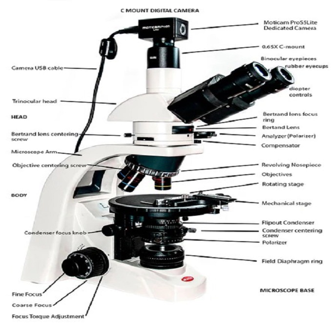
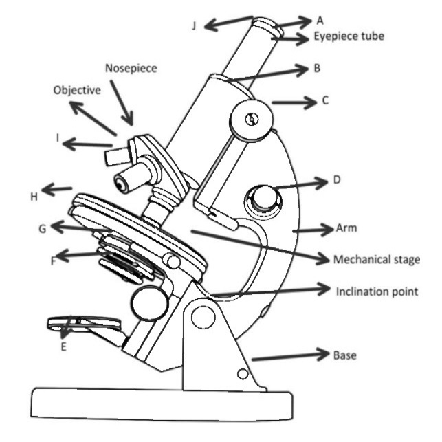
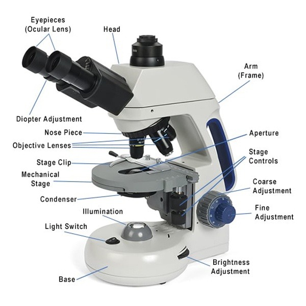
The head, or body tube, is the upper part of the microscope. It holds the ocular lenses, or eyepieces, and connects to the objective lenses by way of the nosepiece. This is where the specimen’s magnified images come into view.
The arm connects the head to the base and is used for carrying the microscope securely. It serves as the main support, ensuring that the sensitive optical parts are held in place, which is vital for maintaining the alignment and focus of the lenses.
The base acts as the foundation, providing stability to prevent the microscope from toppling over. It houses important components, such as the illuminator, which is crucial for projecting light through the specimen. This solid footing is essential as it bears the entire weight of the microscope.
Together, the head, arm, and base form the skeleton that supports all other microscope labeled parts. Recognizing these components is the first step in mastering the use of this indispensable scientific tool.
Exploring the Optical System of a Microscope
The optical system is the microscope’s heart. It makes tiny objects appear large and clear. Let’s explore its key parts.
Eyepiece, Objective Lenses, and Nosepiece
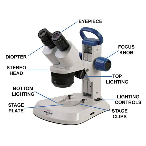
The eyepiece, also known as the ocular lens, is where you peer through to see the magnified image. It usually has a 10x or 15x power. For a closer look, the objective lenses step in. They sit on the nosepiece, a revolving part that lets you switch between lenses of different magnifications. Standard microscopes have usually 4x, 10x, 40x, and 100x lenses. Together, the eyepiece and objective lenses amplify tiny details for your eyes. This combination allows microscopes to reveal a hidden world, otherwise invisible to the naked eye.
The Roles of Focusing Mechanisms
Every microscope labeled needs to bring the viewed object into clear focus. This is where the focusing mechanisms come into play, crucial for fine-tuning the sharpness of the image.
Coarse and Fine Adjustment Knobs
For initial focus, use the coarse adjustment knobs. They move the stage up or down at a greater speed, ensuring that the object comes into the viewer’s field quickly. These larger knobs suit low-power objectives. Fine adjustment knobs are for detailed focus. They adjust the stage height slowly, allowing precise control over the image clarity. This is essential when using high-power lenses, where even slight adjustments can significantly impact the image quality.
Together, these knobs work in unison, ensuring the user can quickly switch from a broad overview to minute details, capturing the intricacies of the specimen under study. Their ease of use is vital for any microscopy work—from students in a classroom setting to professionals in high-stakes research environments.
The Stage: Platform for Specimen Placement
The stage is a crucial part of the microscope. It’s the flat platform where we place the specimen slide for examination. Its design allows for the secure positioning and manipulation of the slide, which is vital for thorough observation of the sample under the lens.
Stage Clips and Mechanical Stage
Stage clips clasp the slide firmly to prevent sliding during focus adjustments. They ensure the specimen stays in place while you switch between different magnification levels. For enhanced control, the mechanical stage comes into play. This advanced feature lets you move the slide smoothly in precise increments. You can adjust the slide’s position with knobs, without touching it. This minimizes disruption and maintains the focus, especially when observing at high magnifications. The mechanical stage is a significant aid in tracking and documenting specific areas of interest on the slide, making it a valued addition to modern microscopes.
Illumination and Imaging Enhancements
To reveal the fine details in specimens, microscopes use an intricate lighting system. This system enhances the clarity and contrast of the image we observe through the eyepiece. Key components contributing to this are the condenser, diaphragm, and illuminator.
Condenser, Diaphragm, and Illuminator
The condenser is a lens that focuses light onto the specimen. It typically sits below the stage and plays a critical role in image clarity. A well-focused beam of light can mean the difference between a vague blur and a sharp image.
Next is the diaphragm, often an adjustable aperture that controls the amount of light reaching the specimen. By changing the diaphragm’s size, one can manipulate light intensity and contrast. This adjustment is crucial for viewing different types of specimens that may require varying light levels.
Finally, the illuminator acts as the microscope’s light source. It’s commonly an LED or halogen bulb positioned near the base. The right amount of illumination is key to viewing a vivid and well-defined specimen. Too much light can wash out the details, while too little can leave you squinting at shadows.
These components together empower the microscope to light up tiny worlds with precision. They make it possible to view structures too small for the eye to see. And, it’s this illumination that sets microscopes apart from mere magnifying glasses. It breathes life into the otherwise invisible and highlights the marvels hidden in plain sight.
Additional Microscope Features
In-depth knowledge of a microscope’s anatomy includes familiarity with its supplementary features. These components may seem minor but are pivotal in the operation of the microscope.
Aperture, Rack Stop, and Light Switch
The aperture is a small opening in the stage. It permits light to travel through the slide, allowing observation of the specimen. Precise control over the light passing through the aperture is essential for high-quality imaging.
The rack stop is a safety mechanism. It prevents the objective lenses from descending too low and potentially crashing into the slide. This feature minimizes the risk of damage both to the slide and the lenses.
Lastly, the light switch is the control button to turn the microscope’s light source on and off. An adjacent control often accompanies it, the brightness adjustment. This allows the user to regulate the intensity of light illuminating the specimen, crucial for enhancing image contrast and details.
These additional features contribute significantly to the microscope’s functionality, ensuring both safety and optimal viewing conditions. Recognizing their roles enhances one’s proficiency in microscopic explorations.
Maintenance and Care of Microscope Parts
Maintaining a microscope in top condition is key to its performance and longevity. Proper care ensures that each part works as intended and that your microscope provides clear, accurate images for years to come. Here are some essential tips for maintaining your microscope labeled parts:
Regular Cleaning
Keep optical surfaces free of dust and oil. Use lens paper or a soft, lint-free cloth dampened with lens cleaning solution. Wipe gently to avoid scratching the lenses. Clean the body and base with a mild, non-abrasive cleaner to keep them looking new.
Handling with Care
Always hold the microscope by its arm and base when carrying it. Avoid touching the lenses with your fingers. Oils from your skin can damage the coatings on the lenses.
Proper Storage
Store your microscope in a dry, dust-free environment. Cover it with a dust cover when not in use. This prevents dust from settling on the parts and helps protect against accidental damage.
Avoiding Moisture
Keep your microscope away from wet or damp areas. Moisture can cause rust and mold, which harm the microscope’s parts. If any part gets wet, dry it immediately with a soft, absorbent cloth.
Regular Inspections
Check your microscope for loose screws or misaligned parts regularly. Make sure all moving parts operate smoothly. Tighten any loose components and consult a professional if you notice any alignment issues.
By following these simple steps, you will keep your microscope working well. This ensures that every magnified glimpse into the microscopic world is as clear and vivid as possible.
Conclusion: Understanding Microscope Anatomy and Function
As we reach the conclusion of our guide, it’s clear that understanding the anatomy and function of a microscope labeled parts is vital. Each piece, from the eyepiece to the base, plays a crucial role in bringing the microscopic world into view. Users must recognize these components to effectively operate the instrument and explore unseen realms. Whether for education, research, or hobby, the knowledge of microscope labeled boosts confidence and skill in using this scientific marvel. Proper care and regular maintenance ensure its longevity and reliability, rewarding users with clear and accurate insights into the minuscule wonders of our world. In sum, the microscope remains an indispensable tool, opening the doors to discovery and enhancing our comprehension of minute details beyond the reach of the naked eye.