Introduction to Microscopes
Microscopes are powerful tools that let us see the tiny world around us. Small things, like cells and microbes, become visible with them. Since their invention, microscopes have been vital for science and medicine.
The Evolution of the Microscope
The first microscope was made in the 16th century. Since then, they have gotten better and better. They started with just one lens and now have complex systems to see even smaller things.
Significance of Microscopes in Science
Microscopes are super important in labs and research. They help scientists see tiny details that the eyes can’t see. This helps them understand diseases, find new cures, and learn more about life itself.
Types and Uses of Microscopes
Microscopes come in different types, each designed for specific purposes. Understanding the various microscope types can help select the right one for each application.
Light Microscopes and Their Applications
Light microscopes are the most common type. They use visible light to illuminate specimens. Schools and simple lab setups often use them. Their main applications include studying cells, simple organisms, and small invertebrates.
Electron Microscopes: TEM and SEM
Electron microscopes, including TEM and SEM, use electrons to create images. They provide much higher resolution than light microscopes. TEM shows the internal structure of cells, while SEM gives detailed surface views.
Stereo Microscopes for Three-Dimensional Observation
Stereo or dissecting microscopes give a 3D view of larger specimens. They are lower in magnification but high in depth perception. They are often used for dissection or examining the surface features of solid objects.
Other Advanced Microscope Types
There are other specialized microscopes as well. These include phase-contrast microscopes for live cells and dark-field microscopes for unstained samples. Each type has its unique role in science and research.
Core Structural Components of a Microscope
The Head or Body Tube Explained
The head, or body tube, is essential to the microscope’s structure. It holds the lenses that magnify the specimen. Located at the top of the microscope, the head connects the eyepiece to the objective lenses. Light reflects within this tube, allowing images to form.
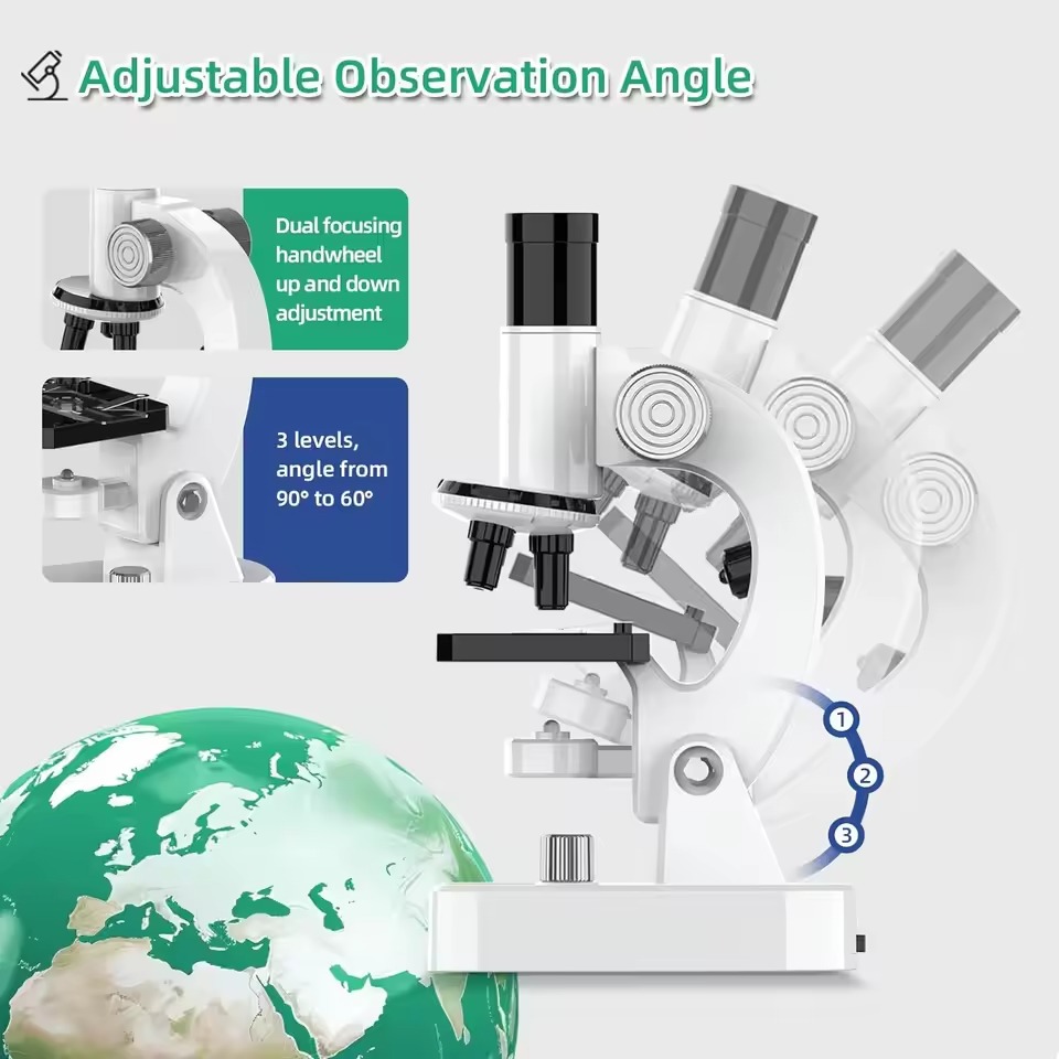
The Functionality of the Microscope Arm
The arm is the part that you use to safely carry the microscope. It supports the instrument’s structure, linking the base and the head. Some high-grade microscopes have arms that can move. This helps in adjusting the head for a better view.
The Supporting Role of the Microscope Base
The base gives the microscope stability. It’s the bottom part of the microscope that houses the light source. All components rest on it, making it critical for the microscope’s balance and support.
Optical Components: The Key to Magnification
Objective Lenses: The Primary Magnifiers
Objective lenses are the main parts of a microscope that increase the size of the specimen image. They sit close to the object being studied. There are often 3 to 4 objective lenses on a microscope, each with a different power for magnifying. These range from low (4X) to high (100X) magnification. The higher the magnification, the closer you can look at small details.
The Role of Eyepieces (Ocular Lenses)
The eyepieces, also known as ocular lenses, are where you look through to see the magnified image. They typically magnify the image by 10X or 15X. The eyepieces work with the objective lenses to make the image larger so you can see tiny things clearly.
The Mechanism of the Nosepiece and Objective Switching
The nosepiece holds the objective lenses and can turn. This allows you to swap between lenses for different magnification powers. To see more detail, you rotate the nosepiece to a higher magnification lens. This is an easy way to switch views without having to move the specimen.
Focusing and Fine-Tuning
The Function of Stage and Stage Controls
Microscopes come with a stage, which is the flat area where we place the slide holding our specimen. This stage can sometimes move. It has knobs which we use to go left, right, up, or down. This helps us find the right part of the specimen to look at. Stage clips may also be present. They grip the slide so it doesn’t move while we are viewing.
Adjustment Knobs: Coarse and Fine Focusing
There are two main knobs for focusing on a microscope: the coarse and fine adjustment knobs. The coarse adjustment knob makes big changes in focus, like when we first put a slide under the microscope. The fine adjustment knob is for small tweaks, for a clearer image especially under high magnification. They work together to get a sharp view.
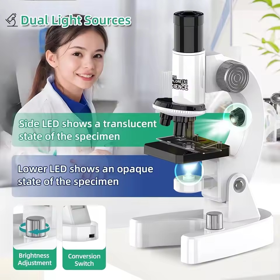
The Condenser and Diaphragm’s Light Control
A condenser sits below the stage. Its job is to focus light onto the specimen. Next to the condenser is the diaphragm. It controls how much light gets through to the specimen. We can adjust it to change the light’s brightness, which affects how we see the specimen. These parts are crucial. They help us get a good image by managing the light just right.
Illumination System in Microscopes
Light Sources and Their Importance
The bright light source of a microscope is key to seeing specimens clearly. It shines light through the specimen, making details visible. Most modern microscopes use LED or halogen bulbs. These provide steady, bright light with the option to adjust brightness.
Without enough light, even with great lenses, we can’t see the tiny world well. Light must be right for the lenses to work. For small specimens, bright light helps show clear images. Every detail and structure becomes easier to explore.
Condensers: Focusing Light on Specimens
The condenser sits under the stage. It’s like a spotlight that focuses light straight on the specimen. It works with the diaphragm to manage light quality and quantity. Adjusting a knob moves the condenser nearer or farther, changing how light hits the specimen.
There’s a sweet spot for seeing things clearly. Too much or too little light can make images blurry. That’s why adjusting the condenser is important. It helps get the light just right for sharp, bright images. With it, we can see tiny structures in great detail.
Conclusion
Microscopes have changed how we study tiny worlds. They help us see and understand micro-level details invisible to the naked eye.
The Impact of Microscopes on Modern Science
Microscopes have hugely impacted science. They are key in biology and medicine, letting us study cells and diseases. Thanks to microscopes, we’ve learned much about life and health.
New medicines and cures are found by looking at tiny microbes. With better microscopes, we can watch cells and bacteria grow. This teaches us how they live and change.
Microscopes have even helped solve crimes by looking at small clues. Very small details at crime scenes can now be seen, leading to key evidence.
Future Advancements in Microscopy Technology
Better microscopy tech is coming. This will let us see smaller details and do new kinds of research.
In the future, microscopes might show us more about atoms and molecules. They could help us create new materials and better drugs. We might even see things we’ve never imagined before.
With new tech, microscopes will work faster and give clearer images. They’ll be easier to use and help more people see the micro world.
Soon, we could have microscopes that connect to computers and share images across the world. This will let scientists work together better. It’s an exciting time for microscopy and science.