Introduction to Cremation Ashes
Cremation transforms the remains of loved ones into ashes. These ashes, when viewed under a microscope, reveal fascinating patterns. Cremation is a process where high heat reduces a body to its basic elements. The result is a fine, powdery substance often referred to as ‘cremains.’ Understanding what happens during cremation helps us appreciate the microscopic world of human ashes. Each particle tells a story. They speak to the life once lived, as well as the journey through fire.
Many don’t know that human ashes under a microscope show unique structures. These structures look different based on various factors. They range from the temperature of cremation to the individual’s elements. In this post, we will explore the ashes left behind. We will look into their composition and what they can tell us. Our aim is to unravel the mystery surrounding the microscopic view of human cremains.
Studying the microscopic patterns in human ashes adds depth to our understanding. It gives a new perspective on what remains after life. As we dive deeper into this topic, keep in mind that these remains once formed a part of someone’s life. In the following sections, we will delve into the detailed analysis of these patterns.

The Basics of Microscopic Analysis
Microscopic analysis is a key tool in examining cremains. Experts use microscopes to magnify human ashes. This reveals intricate details not visible to the naked eye. The process begins with collecting a small sample of ashes. These are then placed on a microscope slide for examination.
Through the microscope, various structures within the ashes become apparent. Each particle may tell a unique story, depending on its shape, size and composition. Patterns emerge that can provide insight into the conditions of cremation. This analysis is critical for a deeper understanding.
The equipment used in microscopic analysis is quite sophisticated. High-powered microscopes are capable of providing a detailed view of particles. These instruments reveal the extraordinary complexity of human ashes under a microscope.
Studying these microscopic patterns requires a careful technique. Analysts must avoid contaminating the sample. They also need to ensure the microscope is correctly calibrated. With precision and care, the hidden world within human ashes unveils itself.
In summary, microscopic analysis allows us to appreciate the intricate patterns found in cremains. It shows us how these remnants encapsulate the history of a human life. In the next sections, we will delve into the structures, composition, and implications of these patterns.
Unveiling the Microscopic Structure of Cremains
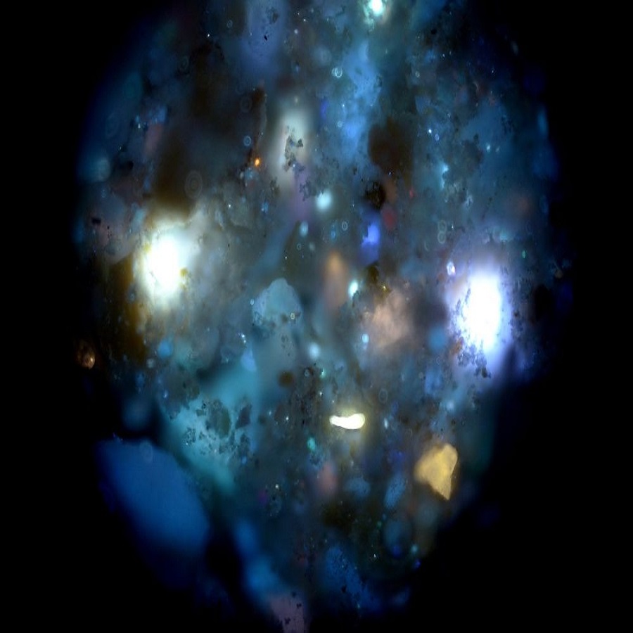
When we place human ashes under a microscope, a hidden universe unveils itself. The structure of cremains is intricate, formed through intense heat during cremation. Microscopic examination shows that what might seem like a uniform powder is actually a collection of diverse particles. The patterns observed are as unique as fingerprints, varying from person to person.
Under the microscope, these structures come to life. We might see crystal-like formations, or even minute irregularities shaped by the life’s journey and the cremation process. These structures often hold minerals that were once part of the human body. They transform under high temperatures, resulting in distinct patterns that are a testament to the individual’s worldly existence.
The microscopic view offers clues about the cremation. Factors such as the furnace’s temperature and the time spent in the flames affect the ashes’ structure. High heat can lead to more fragmented, finer particles, while less intense conditions may leave larger, more discernible shapes. By examining these patterns, we learn how each individual’s physical composition responds to the flames.
The delicate process of uncovering the structure of cremains under a microscope requires steady hands and a keen eye. Specialists carefully place the tiny sample onto a slide, scanning through the lens to explore every corner of this microcosm. The intricate patterns tell a story — one of life, transformation, and the finality of cremation. As we navigate through the following sections, we will delve deeper into the composition and significance of these fascinating structures.
Understanding the Elemental Composition
When observing human ashes under a microscope, we delve into their elemental makeup. The elemental composition reveals the core building blocks of the human body. Ashes contain primarily calcium and phosphorus. These elements remain after the organic matter has burnt away. Magnesium, sodium, and potassium also appear in smaller quantities. These elements link directly to the bones and soft tissues of the body.
The heat during cremation alters the elemental balance. This can cause some elements to become more pronounced in the ashes. Trace amounts of other minerals found in the body may still persist. These can include iron, sulfur, and sometimes heavier metals, depending on prior medical treatments or lifestyle factors.
The presence and ratio of these elements can vary. It often depends on the individual’s diet, age, and health. For instance, younger individuals might have different elemental ratios than older adults. Studying these variances gives insight into the life of the deceased.
Each element leaves behind a unique signature. This signature is visible under a microscope as distinct patterns or colors. Analysts can identify the elements by their signature shapes and colors. Their specific arrangements and how they interact with each other are noteworthy.
In summary, analyzing the elemental composition is crucial. It provides a deeper understanding of the transformation from life to ashes. It shows how the cremation process preserves the trace of a person’s physical existence. In the next sections, we will explore even further details, like particle size, shape, and color variations.
The Significance of Particle Size and Shape
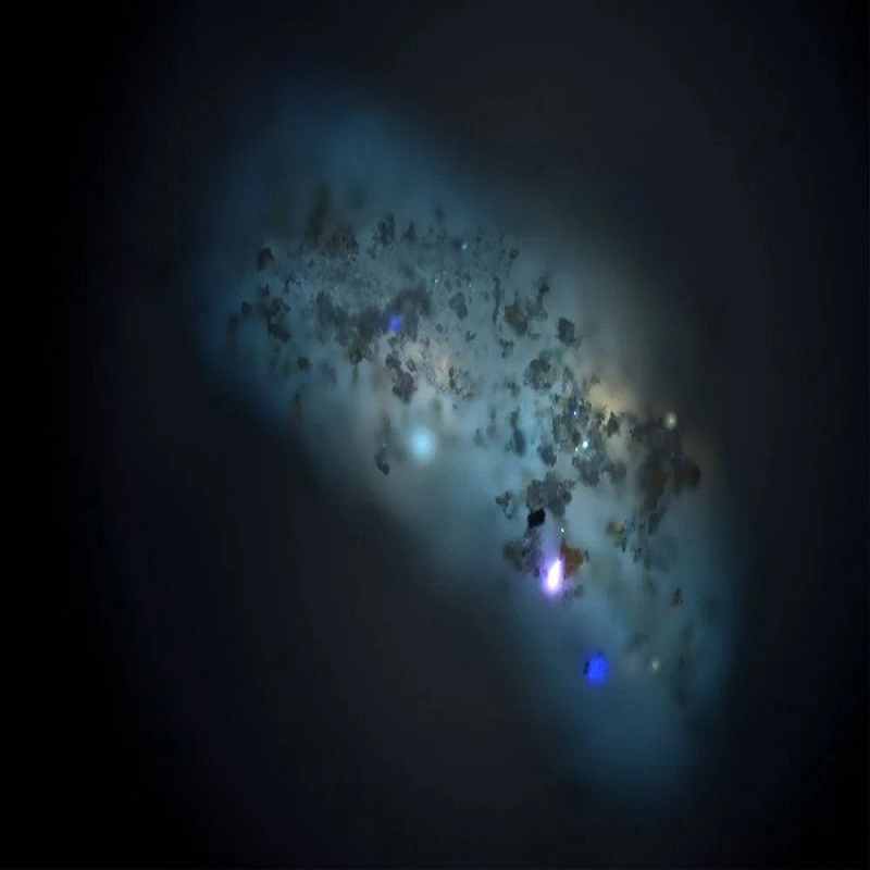
When we examine human ashes under a microscope, particle size and shape stand out. These features are crucial. They can tell us a lot about the cremation process. Small particles often result from very high heat. This intensity causes the ashes to break down more completely. Larger particles may indicate a less intense cremation temperature.
Shape plays a role too. It hints at the history of the ashes. Irregular shapes could suggest less uniform heat during cremation. In contrast, more uniform shapes might show steady heat exposure. Researchers look at these differences to understand the cremation.
Particle size and shape affect how ashes spread. Small, fine particles drift further when scattered. Larger particles are more likely to stay closer to where they are released. Loved ones might consider this when choosing a final resting place for the ashes.
In forensic cases, particle size and shape are vital clues. They can help identify if the remains are human and can even hint at environmental factors at the time of cremation. Experts can sometimes tell if the body was moved or disturbed after death.
To sum up, the particle size and shape in cremains have a significant story to tell. They provide a deeper view into the past of the ashes. They also guide families in making decisions about memorial practices. As we move to the next section, we will explore how color variations further add to this story.
Exploring the Color Variations of Ashes
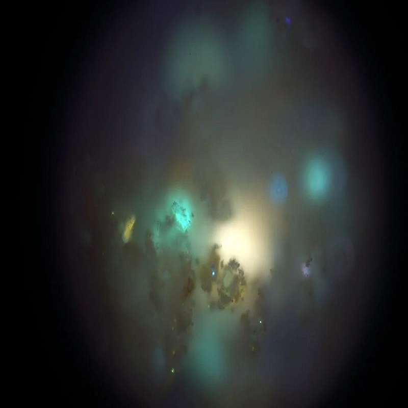
When we examine human ashes under a microscope, the range of colors is remarkable. Each hue reflects a different element’s presence in the body. Lighter shades often point to a higher calcium content, a major part of our bones. Ashes may also show dark specks, where carbon or iron are present.
These varied colors come from the intense heat during cremation. Temperatures change the way elements look, even at a micro level. Calcium might show up as white flecks, while sodium could give a subtle blue tint. The mix of these colors can tell a story about the person’s life, like their diet or health.
To see these colors, experts place a tiny bit of ashes on a slide. Through the microscope, a burst of colors appears. Each particle, with its unique shade, adds to the beauty of the whole sample. It’s like looking at a painting, but every speck is a part of someone’s life story.
The hues also reflect how bones and tissues turn into ashes. Hard tissues like bones leave behind a lot of whites and grays. Soft tissues might result in darker areas, as they hold more carbon.
In essence, color variation in human ashes exposes our body’s chemistry. It gives a visual dimension to what’s left after life fades. Next, we’ll discuss how the cremation temperature impacts these colors and patterns.
The Impact of Cremation Temperature on Ashes
The cremation temperature plays a pivotal role in determining the final appearance of human ashes under a microscope. High temperatures typically cause ashes to fragment into finer particles, reflecting a more thorough breakdown of bone and tissue. With less intense heat, the cremains might retain larger, more identifiable particles.
Cremation temperatures vary from facility to facility and depend on equipment and protocols. A standard range lies between 1400 to 1800 degrees Fahrenheit. Within this spectrum, subtle alterations in temperature can lead to noticeable changes in both particle size and coloration.
At the higher end of this range, remains are subjected to intense heat that often results in lighter-colored ashes. These often display a powdery consistency and a high degree of disintegration. At lower temperatures, one might observe cremains with more varied colors and larger, more distinct structures.
The color spectrum can shift based on temperature as well. Extremely high temperatures have the potential to burn away more organic material, sometimes leaving behind a purer white from calcined bones. Conversely, moderate cremation temperatures might not consume all organic material, resulting in a broader array of colors including grays and darker tones.
By examining the consequences of cremation temperature on human ashes, it becomes clear how closely tied the physical properties are to the process itself. These insights can help us understand the transformation that occurs during cremation and offer a unique window into the journey from life to the legacy left in ashes.
Potential Applications in Forensic Science
The detailed study of human ashes under a microscope extends beyond mere curiosity. It serves significant roles in forensic science. By analyzing the microscopic patterns, forensic experts can gather vital clues. These may relate to the identity of the deceased or circumstances surrounding their cremation.
Forensic teams can compare known samples to those found at a scene. This can lead to confirmations of a person’s presence or reveal if remains have been tampered with. Particle size and shape, along with color variations, aid in such determinations. For example, irregularly shaped particles may hint at uneven heating or possible movement of the body post-cremation.
Moreover, the elemental composition of cremains can be revealing. They can suggest the deceased’s diet, health, or even exposure to certain environments. Such detailed analyses can even draw conclusions about the temperature of cremation. This is crucial in investigations that deal with improper or illegal cremation practices.
In essence, while often overlooked, the study of human ashes under a microscope is powerful. It can solve cases and bring closure to grieving families. As technology and methods improve, its applications in forensic science will likely expand even more.
Conclusion: The Mysterious Beauty of Ashes Up Close
Viewing human ashes under a microscope opens a world of wonder. It unveils the beauty in the ashes left behind. Such analysis transforms our view of cremation. It’s more than a process. It becomes a story of transformation from the physical to the microscopic. Through this blog, we have discovered just how much can be learned. From shapes to colors, every particle has significance.
The microscopic patterns in cremains tell rich tales. Tales of individual existence and the journey through cremation. These patterns are unique to each person, like a microcosm of their life. They capture a final snapshot. It’s a scientifically beautiful tribute to what once was.
In forensic science, this study is useful. It unravels mysteries and serves justice. It turns ashes into evidence. And for the families, it might offer a deeper connection. In their grief, seeing these patterns could provide a new layer of meaning.
The blog journey has shown us the fine details of cremains. It’s highlighted the science and the significance. Beyond the technical, it encourages a new appreciation. The dance of elements and conditions during cremation is almost an art. It’s a poignant reminder of life’s fragility and complexity.
In closing, the microscopic world of human ashes tells us so much. It confirms that even in ashes, beauty endures. It reminds us that, even at the end, there’s much to discover. And perhaps most importantly, it shows how life’s essence lingers. It lingers in the finest grains, waiting for someone to look closer and see the mysterious beauty of ashes up close.