The Basics of Viral Structure and Function
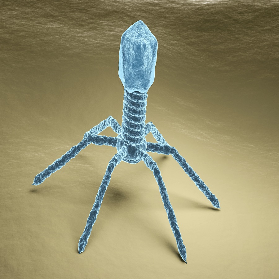
Understanding viruses begins with a grasp of their basic structure and function. At their simplest, viruses consist of genetic material wrapped in a protein coat called a capsid. This structure can vary among different virus types, with some containing additional layers, like an envelope of lipids that surrounds the capsid. The genetic material within can be DNA or RNA, single or double-stranded.
The primary function of a virus is to replicate itself inside the host cell. To do this, a virus must first attach to a host cell using specific proteins on its surface. Once inside, it hijacks the host cell’s machinery to produce new virus particles. These new viruses can then go on to infect more cells, continuing the cycle.
Viruses are incredibly efficient at what they do but they’re not considered living organisms since they cannot reproduce or carry out metabolic processes on their own. Their activities depend entirely on their ability to invade and take over living cells.
By studying a virus under microscope, researchers can learn about these basic components and functions in detail. This knowledge helps in developing vaccines, understanding how viruses evolve, and how they interact with their hosts.
Microscopy Techniques for Virus Observation
To observe viruses, scientists use several microscopy techniques. These techniques reveal the intricate details of viral structure.
One common method is light microscopy. This technique uses visible light and can magnify up to 1,500 times. However, light microscopy has its limits. Most viruses are too small to see with this method alone. They are smaller than the wavelength of visible light, making them difficult to image.
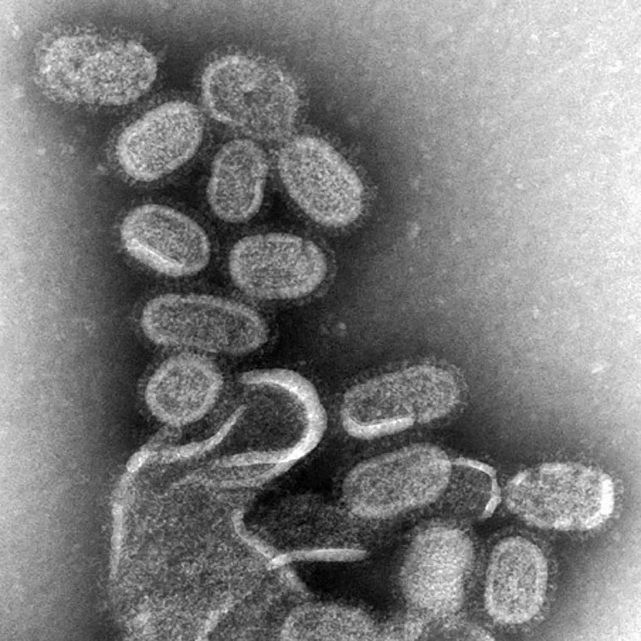
For higher magnification, scientists turn to electron microscopy. Electron microscopes use beams of electrons instead of light. This allows them to achieve much greater magnification and resolution. Transmission electron microscopy, or TEM, can magnify viral particles over 100,000 times. Scanning electron microscopy, or SEM, offers 3D views of virus surfaces.
Fluorescence microscopy is another useful technique. It involves tagging viruses with fluorescent markers. These markers glow under certain lights, helping to differentiate viruses from other cellular components.
Each of these techniques can show different aspects of viral structure and infection processes. They are crucial tools for virologists in understanding how viruses function and interact with host cells.
The Role of Electron Microscopy in Virology
Electron microscopy has revolutionized the study of virology. It allows an in-depth look at viruses that was once impossible with traditional light microscopy. The high resolution of electron microscopes reveals the intricate details of a virus under microscope that are not visible with other techniques.
Transmission electron microscopy (TEM) and scanning electron microscopy (SEM) are the two main types used in virology. TEM offers views inside the virus, showing the arrangement of genetic material and proteins. SEM gives us 3D images of a virus’s surface. This lets us see how they interact with host cells.
Scientists use these powerful tools to understand a virus’s life cycle. They can watch a virus as it attaches to and enters a host cell. They observe how a virus hijacks the cell’s machinery to replicate itself. Each stage of infection becomes clearer under the powerful lens of an electron microscope.
Electron microscopy also aids in vaccine development. By seeing the virus’s structure, scientists can identify parts of the virus for target vaccines. This knowledge leads to safer and more effective vaccines.
In short, electron microscopy provides a crucial window into the world of viruses. It is a key tool in uncovering the secrets of viral behavior and structure. With it, virologists can develop therapies and vaccines to fight viral infections.
Recent Discoveries About Viruses Through Microscopy
Microscopy has unveiled new insights into virus behavior and structure. Recent studies using advanced microscopy techniques have made significant discoveries. For instance, researchers have observed the exact moment a virus pierces a cell membrane. This is critical for understanding infection mechanisms.
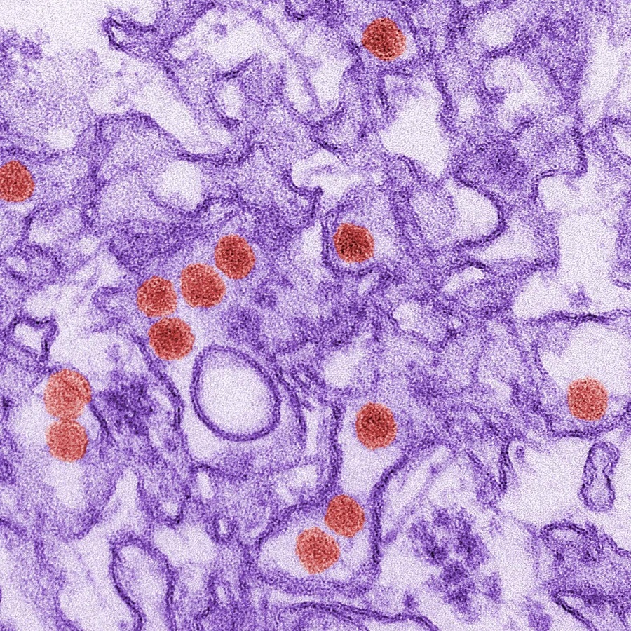
The use of cryo-electron microscopy (cryo-EM) has surged in popularity. Cryo-EM allows scientists to see viruses at atomic resolution in a near-natural state. One groundbreaking discovery was the detailed structure of the Zika virus. This understanding is vital for vaccine development.
Another major development is the visualization of virus assembly. Microscopy now shows us how viral components come together within a host cell. This was previously a mystery due to the complex nature of the process.
Moreover, the observation of viral infection in real-time has been a leap forward. Time-lapse imaging techniques have provided a dynamic view of viral entry and replication. This helps in predicting virus spread and potential outbreaks.
Imaging of virus-host interactions has also revealed surprising details. It showed the various strategies viruses use to evade the immune system. These findings are changing the way we think about antiviral therapies.
In summary, microscopy has become an indispensable tool in virus research. It allows for observations that lead to direct and actionable knowledge. This greatly aids in the fight against viral diseases. Each discovery through the lens of a microscope is a step closer to curbing the impact of viruses on human health.
Common Challenges in Imaging Viruses
Imaging viruses presents numerous challenges for scientists. The minute size of viruses is a primary hurdle. They are often smaller than the wavelength of visible light. This makes them difficult to see using standard light microscopy. As a result, more sophisticated techniques, like electron microscopy, become essential.
Another issue is keeping viruses intact during observation. Sample preparation for electron microscopy may alter or damage delicate viral structures. Care is required to preserve the natural state of the virus. Cryo-electron microscopy helps by freezing viruses quickly. This technique maintains their shape and form for study.
Viruses also vary greatly in shape and size. This diversity means that no one imaging technique is perfect for all viruses. Researchers must choose the right method for each virus.
Another challenge is the dynamic nature of viruses. Capturing them in action, as they infect cells, requires precise timing and methods. Time-lapse imaging and other real-time techniques are vital. They must be used carefully to observe these quick processes.
Lastly, the high cost and complexity of advanced microscopy limit access. Not all research facilities can afford such equipment. This hampers the spread of knowledge and the speed of scientific advancement.
These challenges require creative solutions and technological advancements. Overcoming them will lead to deeper insights into viral behavior and structure.
Advancements in Virus Visualization
Recent years have seen notable progress in virus visualization. Advanced techniques are shining new light on the unseen world of viruses. These improvements are essential for greater precision in research and treatment.
One key development is the refinement of cryo-electron microscopy (cryo-EM). Cryo-EM freezes viruses in their tracks, allowing examination at atomic levels. We now see viruses in states close to their natural form. This detail-rich view is a game-changer for virology.
Super-resolution microscopy is another leap ahead. It breaks the limits of light microscopy by improving resolution. It allows us to see particles even smaller than light wavelengths. This method brings the subtle structures of viruses into sharp focus.
Time-lapse imaging techniques have also improved. They let scientists watch viruses as they infect cells in real time. This dynamic view provides insights into how viruses move and multiply. Such understanding is key for developing strategies to block infections.
Artificial intelligence (AI) is playing a growing role in virus visualization as well. AI algorithms process vast amounts of data from images. They help decipher complex patterns that humans might miss. This approach speeds up research and opens new paths for discoveries.
To sum up, advancements in visualization techniques allow us to see viruses like never before. From cryo-EM’s atomic-level shots to AI’s data crunching, these tools are invaluable. They not only push forward our understanding but also aid in creating better treatments.
The Importance of Understanding Viruses in the Medical Field
Understanding viruses plays a critical role in the medical field. Accurate knowledge of viruses helps prevent diseases. It guides the creation of effective vaccines and treatments. One classic example is the development of the flu vaccine. Researchers study the flu virus under a microscope every year. They track how it changes from the previous season. This helps them forecast which flu strains will be most common.
Virus observation also aids in rapid response to outbreaks. Knowing how a virus moves between cells assists in containment efforts. This was evident in the recent global efforts to understand and control COVID-19. Microscopes allowed researchers to study the virus closely. They learned how it infects humans and how it spreads. This knowledge was vital in developing testing kits and safety protocols.
In hospital settings, virus understanding is key to patient care. It helps doctors diagnose infections more accurately. They can choose the most effective treatment options. In cases of unknown illnesses, virus observation can identify new diseases. This leads to quick development of new medical responses.
Studying viruses under the microscope further contributes to public health education. It provides solid evidence of how viruses function. This can improve vaccine acceptance and adherence to public health guidelines. Educating the public about viruses reduces fear and misinformation. It promotes healthy behaviors that prevent virus spread.
In summary, a deep understanding of viruses is fundamental. It safeguards human health on many levels. From developing vaccines to guiding outbreak responses, microscopy offers an invaluable look into the viral world. This understanding is crucial in protecting public health.
The Future of Virus Research and Microscopy Techniques
The path ahead in virus research is promising due to advancements in microscopy techniques. Looking to the future, scientists anticipate more breakthroughs that will deepen our understanding of viruses and enhance medical research. As we continue to confront new viral threats, the evolution of microscopy will be pivotal.
Upcoming trends in the field include the integration of new imaging modalities with artificial intelligence (AI) and machine learning (ML). These technologies will likely make virus analysis faster and more accurate. AI can process complex images quickly, identifying patterns that may elude human researchers. This could expedite vaccine and antiviral drug development.
Another anticipated advancement is the improvement of in situ techniques. These allow the observation of viruses within their natural or living environments. Researchers will be able to see how viruses interact with host cells in real time. This might uncover previously hidden aspects of viral life cycles.
Portable microscopy devices are also on the horizon. They could bring virus visualization capabilities to remote or resource-limited areas. This would be critical for early detection and response to viral outbreaks in under-served communities.
Collaboration across disciplines will further enhance virus research. Combining insights from virology, immunology, and molecular biology with cutting-edge microscopy could yield holistic views of viral infection and immunity mechanisms.
Finally, ethical considerations will play an increasing role in virus research. As technology allows us to see and manipulate viruses at more fundamental levels, the scientific community must address the potential implications responsibly.
In summary, the future of virus research is closely tied to the evolving landscape of microscopy techniques. These technologies promise to unveil the intricate details of viruses under the microscope, leading to better understanding and control of viral diseases.