Introduction to Plant Microscopy
The microscopic world of plants holds many secrets. Viewing grass under a microscope reveals intricate structures invisible to the naked eye. Scientists and hobbyists use plant microscopy to study these details. It offers insights into how plants function and interact with their environment.
What is Plant Microscopy?
Plant microscopy is examining plant parts at the cellular level using a microscope. This practice allows us to see the composition and arrangement of cells. It also helps in understanding plant biology and the role of various cells.
Why Study Grass Under a Microscope?
Observing grass under microscope can teach us much about plant health. It can help with disease diagnosis in crops. This study also contributes to genetic research and breeding programs.
Tools Used in Plant Microscopy
Several tools aid in plant microscopy. A compound microscope is the main one used for viewing grass cells. Others include slides, coverslips, stains, and microtome for sectioning samples.
Understanding Microscopy Terms
Familiarity with some basic terms is important. Magnification refers to how much larger an object appears. Resolution is the clarity of the magnified image. Staining is a technique used to enhance contrast in microscopic images.
In the following sections, we’ll delve deeper into viewing grass under the microscope. We’ll explore how to prepare samples, identify grass species, and understand grass anatomy at high magnification. Stay tuned for a fascinating journey into the microscopic world of grass.

The Basic Structure of Grass Cells
When we observe grass under a microscope, the basic cellular structure becomes clear. Each cell is a building block of the grass blade. Cells work together to perform functions necessary for the plant’s survival and growth. Understanding the basic structure of these cells is key to studying grass at the microscopic level.
Grass cells are primarily composed of a rigid cell wall that gives the plant structure and support. Inside the cell wall is the cell membrane, which regulates the entry and exit of substances. The cytoplasm, a gel-like substance, fills the majority of the cell’s interior. Here, various organelles perform essential biological processes.
The nucleus, found in the cell’s center, is crucial as it contains DNA and controls cell activities. Chloroplasts are another vital component, especially visible when looking at grass under a microscope. These organelles are responsible for photosynthesis, which converts light energy into chemical energy the plant can use.
Vacuoles, large storage organelles, are also prominent in grass cells. They store nutrients and waste products and help maintain water balance. Additionally, tiny structures called mitochondria provide the energy needed for the cell’s metabolism by converting sugar into ATP (adenosine triphosphate).
By understanding the composition and function of these cellular structures, researchers can gain insights into how grass grows, reproduces, and responds to environmental stress. The basic structure is the same across different species. However, minute differences aid in identifying various types of grasses under microscopic examination.
In the next sections, we’ll discuss how to prepare these samples for viewing and how to identify different grass species based on their cellular features.
Preparing Grass Samples for Microscopic Examination
Preparing grass samples for microscopic examination is a meticulous process that requires precision. The quality of your preparation greatly impacts the clarity and detail of the microscopic images you will observe. Here are the essential steps you’ll need to take to get your grass samples ready for observation under a microscope.
Cleaning and Selection: Start by carefully selecting the grass blades you wish to examine. Make sure they are clean and free from dirt or debris. This ensures that the microscopic images are not obscured.
Sectioning: Using a microtome, slice thin sections of the grass. Thin sections are crucial as they allow light to pass through, enabling clearer observation of cellular structures.
Fixation: Fix the sections onto glass slides. This process preserves the cellular structure and prevents degradation over time.
Staining: Apply stains to the samples. Stains like iodine or methylene blue can help differentiate between cellular components when placed under a microscope.
Mounting: Cover the stained section with a coverslip carefully to avoid air bubbles. The samples are then ready for examination with a compound microscope.
By following these steps, you enhance your ability to see the minute details present in the cells of the grass under the microscope. It allows for a more accurate study and understanding of grass anatomy at the cellular level.
Microscopic Identification of Grass Species
Identifying grass species under a microscope is an intricate task. Scientists rely on cellular characteristics to distinguish between different types. When viewing grass under a microscope, you’ll notice variations in cell shape, size, and arrangement that are unique to each species. Staining often reveals patterns within the walls of cells that can serve as identifiers.
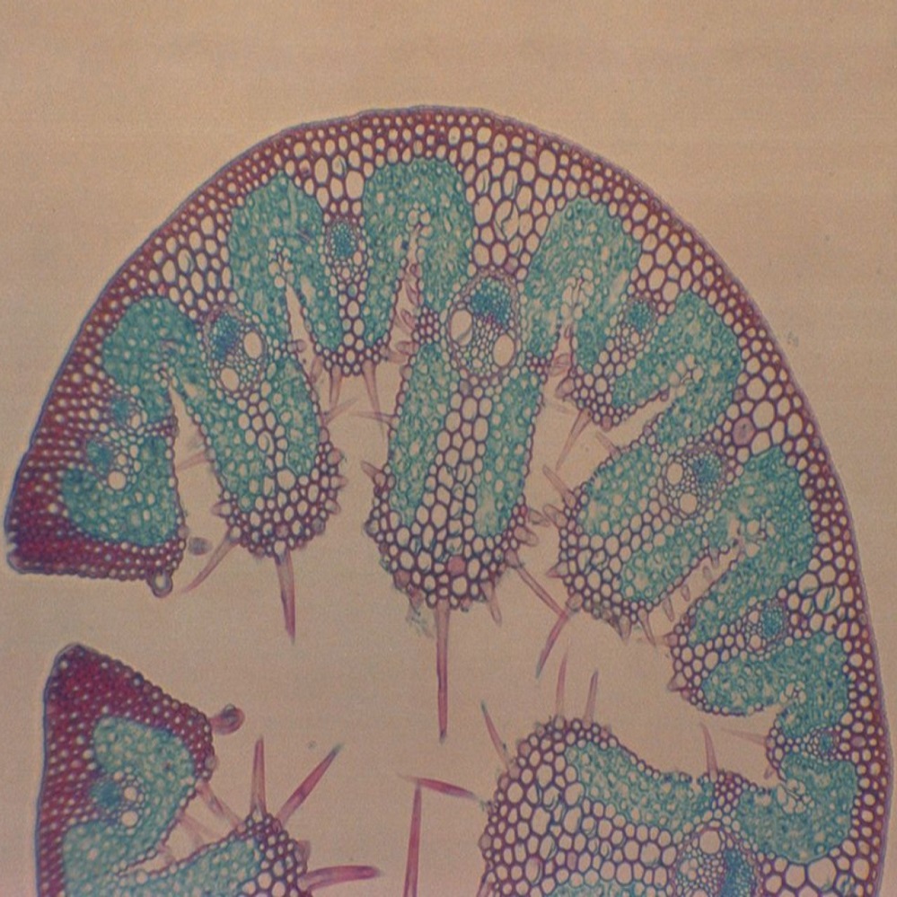
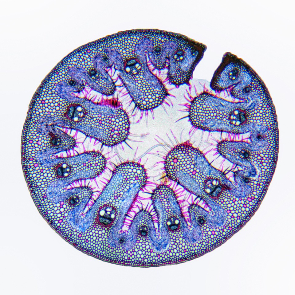
To differentiate species, focus on the structure of leaf blades. Look for the Kranz anatomy, typical in C4 plants like corn, which appears as a wreath-like arrangement of cells around the vascular bundles. Another aspect is the size and number of chloroplasts, which can vary among species.
For more detailed identification, consult a botanical reference that lists cellular features of common grasses. It helps correlate your microscopic observations with known grass species data. This approach ensures accurate identification necessary for ecological studies, landscape management, and agricultural applications.
Through careful examination and comparison with reference materials, researchers can confidently identify grass specimens down to the species level using plant microscopy techniques.

Common Cellular Features in Different Grass Types
While grass species may vary, they share some common cellular features that are observable when we study grass under a microscope. Knowing these features helps scientists and hobbyists alike to understand what makes grasses similar and what distinguishes them. Here are some cellular characteristics that are typically found across different types of grass:
- Cell Walls: Grass cells have strong, rigid cell walls that provide support and give shape to the plant.
- Chloroplasts: These organelles are prominent in grass cells and are essential for the process of photosynthesis.
- Vacuoles: Large vacuoles are present in grass cells, playing a key role in nutrient storage and water regulation within the cell.
- Inter-cellular Spaces: These spaces between cells facilitate the exchange of gases and transport of water and nutrients.
- Plasmodesmata: These are small tubes that connect plant cells, allowing communication and transport of substances between them.
Despite these commonalities, genetic and environmental factors lead to adaptations in the cellular structure. These adaptations are part of what allows for the identification of different species when examining grass under a microscope. In addition to shared features, each grass type has its unique cellular signatures that can be used to tell them apart.
Next, we’ll examine grass anatomy at higher magnification to understand the intricate details that distinguish one species from another. Stay tuned as we continue our microscopic exploration of the hidden world of grass.
Grass Anatomy at High Magnification
When we zoom in on grass under a microscope, a new world of fine details emerges. At high magnification, you can observe characteristics that are not discernible at lower magnifications.
Epidermal Cells: The outer layer of cells, known as the epidermis, serves as a protective barrier. You’ll notice these cells are tightly packed without spaces.
Stomata: These are tiny openings on the leaf surface that control gas exchange. They are surrounded by guard cells that open and close the pore.
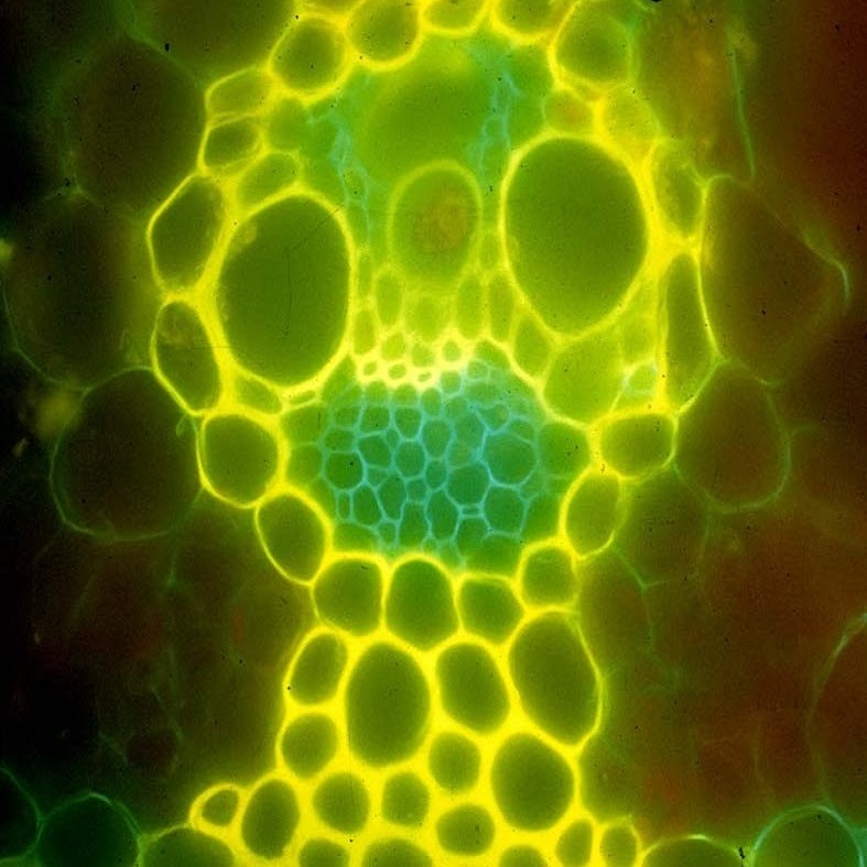
Kranz Anatomy: Seen in certain species, this special structure consists of concentric circles of cells around vascular bundles. It’s a marker for C4 photosynthetic pathways.
Bulliform Cells: These large, bubble-like cells help the leaf to fold during drought, reducing water loss. They’re a distinctive feature under high power.
Vascular Bundles: Look for the xylem and phloem, which transport water, nutrients, and sugars. They form a network throughout the grass leaf.
Subsidiary Cells: Surrounding the stomata, these help in opening and closing of the stomatal pore, playing a significant role in the plant’s breathing process.
By examining grass at high magnification, researchers can learn more about these structures and how they contribute to the life of the plant. It’s like unlocking the secrets to grass’s resilience and adaptability to its environment. This detailed study under a microscope reveals the intricate design and complexity of grass, an everyday plant that’s often overlooked. Each cellular component we’ve discussed plays an essential role in the overall functioning and survival of the grass species.
Photomicrography Techniques for Grass
Learning photomicrography techniques is vital when exploring grass under a microscope. These methods help capture detailed images of grass cells. Improving the clarity and quality of photos can provide valuable data for research. Here’s how to fine-tune your photomicrography skills for grass specimens:
Camera and Microscope Integration: Use a specialized camera designed to attach to the microscope. This ensures high-resolution images that reveal fine details.
Lighting Considerations: Proper lighting is crucial. Adjust the microscope’s light source to avoid glare and shadows. Even illumination brings out the best in cellular structures.
Image Stacking: Some cells have depth. Layer multiple images at different focus points using software. This technique creates a clear, comprehensive view of the entire cell structure.
Adjusting Magnification: Start with a lower magnification to find your subject. Gradually increase magnification for finer details, keeping the image sharp.
Fine Focus: Use the microscope’s fine focus knob for precision. A crisp focus is key to capturing the intricate patterns of cell walls and organelles.
Image Editing Software: Use software to enhance image contrast and sharpness. However, maintain the authenticity of the original picture.
Annotation: Label your images with magnification levels, species names, and cell parts. Clear labels help viewers understand what they’re observing.
Steady Mounting: Reduce camera shake with a stable mount. Vibrationscan blur the details you’re trying to capture.
Photomicrography turns the invisible into something amazing to behold. Practice these techniques to master the art of capturing grass cells under the microscope. Your images can support scientific discovery and appreciation of plant life.

Applications of Grass Microscopy in Science and Agriculture
Exploring grass under a microscope has practical applications in both science and agriculture. Here are key areas where plant microscopy makes a significant impact:
Disease Diagnosis: Microscopes help detect early signs of disease in grasses. Recognizing the cellular changes can prevent widespread crop damage.
Breeding Programs: By studying grass cells, scientists understand traits for breeding. They select varieties with desirable features, such as drought resistance.
Soil Health Monitoring: Observing root structures helps assess soil conditions. Healthy root cells indicate good soil health, essential for productive farms.
Pesticide Research: Microscopy aids in examining how grass cells react to pesticides. This knowledge guides the development of safer, more effective products.
Genetic Research: Looking at grass on a cellular level contributes to genetic studies. It helps identify genes linked to growth and stress resistance.
Ecological Studies: Microscopes are used to study how grasses adapt to their environment. Learning about these adaptations assists in ecosystem management.
Educational Tool: Grass microscopy serves as a teaching aid. It provides students with an up-close look at plant biology and ecology.
The study of grass under the microscope is not just for curiosity. It drives innovation in agriculture, enriches scientific understanding, and helps manage the ecosystem. The knowledge gained through this meticulous process aids in creating a sustainable future for food production and environmental conservation.