The Fundamentals of Fluorescence Microscopy
Fluorescence microscopy is a vital imaging tool in scientific research. This technique hinges on the emission of light by a substance, or fluorophore, that has absorbed light or other electromagnetic radiation. Essentially, the fluorescence microscope allows scientists to light up specific parts of a cell or molecule with fluorescent dyes. These parts become visible against a dark background, creating a high-contrast image.
To begin with, the basic principles of fluorescence microscopy involve excitation and emission. When a fluorophore absorbs light at a certain wavelength, it gets excited. Shortly after, it emits light at a longer wavelength as it returns to a lower energy state. This emitted light is then captured to form an image.
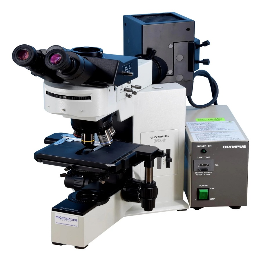
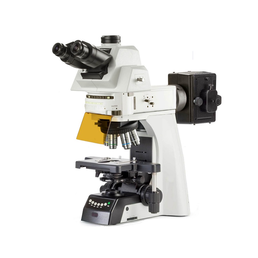
Moreover, the core equipment includes the light source, usually a laser or arc-lamp, to provide the excitation light. The optical filters are critical—they ensure that only the desired wavelength reaches the sample and that only emitted light is detected. The dichroic mirror plays a key role here, reflecting the excitation light toward the sample while letting the emission light pass through.
The detector, which is often a camera or photomultiplier tube, captures the emitted light. The final image reveals vibrant details that are often invisible to the naked eye or with traditional brightfield microscopes. In summary, understanding these fundamentals is the first step in mastering the use of a fluorescence microscope for various research applications.

Key Components and Equipment
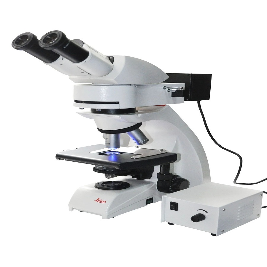
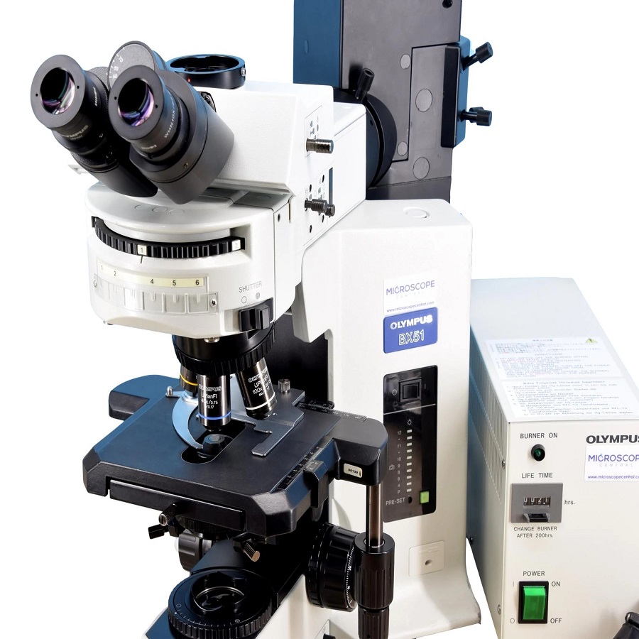
When diving deeper into the workings of a fluorescence microscope, it’s essential to understand the various components that make it function effectively. These components not only include the light source, optical filters, and the detector that were previously mentioned but extend to other critical pieces as well.
The precise alignment of these parts ensures the highest quality images. Condensers focus the light from the light source onto the sample. The objective lens gathers the emitted light and directs it to the detector. Stage controls allow precise movement and positioning of the sample. Finally, the software used for image capturing and analysis is instrumental in extracting meaningful data from the images.
Types of Fluorophores
Fluorophores are the molecules that, upon excitation, emit light and are central to fluorescence microscopy. There are several types of fluorophores used, each with specific properties suited for different applications. Organic dyes like fluorescein and rhodamine have been used extensively due to their brightness and variety of colors. Phosphorescent probes, which emit light over a longer period, allow for time-gated imaging to reduce background noise.
Moreover, fluorescent proteins such as GFP (Green Fluorescent Protein) have revolutionized the field by allowing live cells and organisms to be labeled directly. Quantum dots also offer a broad excitation range and resistance to photobleaching. The choice of fluorophore depends largely on the specifics of the research, such as the required brightness, photostability, and the particular wavelengths of excitation and emission needed.
The Role of Filters in Fluorescence Imaging
Filters in fluorescence microscopy are not merely accessories—they are crucial for the clarity and contrast of the resulting image. Excitation filters control the wavelength of light that reaches the fluorophore, ensuring precise excitation. Emission filters then select the wavelength of the emitted light that reaches the detector. This selection eliminates any unwanted wavelengths, thereby enhancing the contrast and quality of the final image.
A dichroic mirror, or beamsplitter, serves as both a mirror and a filter. It reflects the shorter, excitation wavelengths towards the sample while allowing the longer, emission wavelengths to pass through to the detector. Selecting the right combination of filters and dichroic mirrors is paramount to achieving optimal imaging results. They must match the fluorophores used to maximize signal and minimize noise, ultimately ensuring that the data gathered is both accurate and reliable.
Advanced Techniques in Fluorescence Microscopy
To advance research further, scientists have developed several advanced techniques in fluorescence microscopy. These methods enable deeper insight into biological processes at the cellular and molecular levels.
Confocal Microscopy
Confocal microscopy adds a new dimension to fluorescence imaging. It uses a laser to excite the fluorophores and a pinhole to eliminate out-of-focus light. This results in sharper images with increased resolution. By taking multiple, thin, optical slices and stacking them, researchers can construct detailed 3D models of specimens.
Multiphoton Microscopy
Multiphoton microscopy is another powerful technique that uses infrared lasers to excite fluorophores. It can image deeper into tissue samples since longer wavelengths penetrate further. This method also reduces photobleaching and photodamage, thus preserving the sample.
Super-Resolution Imaging
Breaking the limits of conventional microscopy, super-resolution imaging techniques, such as STED and PALM, achieve resolution beyond the diffraction limit of light. They allow for visualization of structures at the nanometer scale. This level of detail was once thought impossible to observe with light microscopy.

Applications of Fluorescence Microscopy in Research
Fluorescence microscopy has transformed research, offering unparalleled insight into the inner workings of cells and tissues. Its applications span diverse fields, shedding light on complex biological processes.
Cellular and Molecular Biology
In cellular and molecular biology, fluorescence microscope techniques reveal cell structures and protein interactions. Scientists tag proteins with fluorophores and track their behavior in live cells. This allows them to observe dynamic processes like cell division, signal transduction, and gene expression in real time.
Neurobiology and Brain Mapping
Neurobiology also benefits from fluorescence microscopy. Researchers visualize neural networks and brain tissues to understand brain function and structure. They study neural activity, track the growth of nerve cells, and map intricate connections within the brain, aiding in the understanding of neurodegenerative diseases.
Pathology and Disease Diagnosis
Pathologists use fluorescence microscopy to diagnose diseases. They examine tissues and cells, looking for abnormalities. Marking diseased cells with specific fluorophores helps in the detection and understanding of cancer, infections, and genetic disorders. This approach enhances the accuracy of diagnoses and guides treatment strategies.
Optimizing Your Fluorescence Microscopy Setup
Successful fluorescence microscopy relies on optimal setup and meticulous sample preparation. These steps are critical to achieve the best possible image clarity and data accuracy. Here, we’ll discuss how to prepare your samples effectively for fluorescence imaging and the importance of regular microscope maintenance.
Sample Preparation
Proper sample preparation is paramount in fluorescence microscopy. Here are key steps to take:
- Fixation: This process stabilizes the structure of your samples. Use chemicals like formaldehyde to preserve cellular components.
- Permeabilization: Apply detergents to make cell membranes permeable. This step allows dyes to enter the cells.
- Staining: Carefully apply fluorophores to target specific molecules or structures.
- Mounting: Place your stained samples onto slides with appropriate mounting media to protect and preserve fluorescence.
By following these steps, you ensure the fluorescence microscope captures the best quality images. This enhances the overall outcome of your research.
Microscope Maintenance
Regular maintenance is essential for consistent performance of fluorescence microscopes. Ensure these practices:
- Clean Lenses: Wipe objective and eyepiece lenses with lens paper to keep images sharp.
- Check Filters: Inspect and clean filters regularly to avoid image degradation.
- Update Software: Keep your imaging software updated to leverage new features and fixes.
- Lamp Care: Monitor and replace the light source as needed to maintain illumination quality.
Attending to these maintenance tasks will extend the lifespan of your equipment and guarantee that it’s operating at peak efficiency. Implementing a routine maintenance schedule prevents unexpected disruptions in your research work.

Overcoming Common Challenges in Fluorescence Imaging
Fluorescence imaging is a complex process. It often faces challenges that can affect the quality of images. Understanding these challenges is crucial for high-quality results. Here we discuss some common issues and offer solutions.
Photobleaching is a major concern in fluorescence microscopy. It occurs when fluorophores lose their ability to emit light. To reduce photobleaching, minimize light exposure. Use anti-fade reagents in mounting media. These steps help preserve fluorescence.
Another issue is background fluorescence. This is unwanted signal that obscures details. To combat this, use proper controls and enhance washing steps post-staining. Optimize filter settings to ensure specificity of the detected signal.
Fading signal during long-term observations can also be challenging. Use stable fluorophores with high photostability. Opt for fluorescent proteins or quantum dots if longer observations are needed.
Misalignment of optical components may distort images. Regularly check and calibrate your equipment. Ensure alignment is precise for the best image quality.
Sample damage from high-intensity light is a risk too. It can alter the sample’s natural state. Employ gentler illumination techniques like LED sources or reduce exposure times.
Finally, fluorophore overlap can lead to incorrect interpretations. Choose fluorophores with distinct emission spectra. Use spectral unmixing software to separate overlapping signals.
By addressing these challenges, your fluorescence microscope will yield better images. This enhances the reliability of your research findings.
Future Trends and Innovations in Fluorescence Microscopy
The field of fluorescence microscopy is always evolving. Innovations and trends push the boundaries of what’s possible. In imaging, this means greater detail, faster processing, and more efficient workflows. Researchers and developers constantly seek new ways to enhance imaging techniques. Let’s delve into two key areas of progress.
Integration with Other Imaging Modalities
Integrating fluorescence microscopy with other imaging methods is a significant trend. This combined approach provides a more complete picture of cellular events. For instance, merging fluorescence with electron microscopy gives high-resolution images. This allows for detailed study of cell structures. Coupled with live-cell imaging, this integration shows dynamic processes within their ultrastructural context. Together, these methods lead to better insights into cellular functions.
Automation and High-Throughput Screening
Automation is changing how we use fluorescence microscopes. Automated stages and focus systems save time and improve consistency. High-throughput screening is another area benefitting from automation. With it, scientists quickly analyze thousands of samples. They use advanced software to process and interpret data. This means faster research and discovery. New drug candidates or genetic markers are found at an impressive speed. Together, automation and high-throughput screening reshape research and development. They allow for vast amounts of data to be sifted through with precision. This opens doors to new findings in fields like genomics and drug discovery.
