Introduction to Cardiac Muscle Tissue
Cardiac muscle tissue is unique in the human body. It makes up the heart’s walls and helps pump blood. It’s different from other muscles in look and function. Cardiac muscle cells are called cardiomyocytes. They are branched and fit tightly together. Under the microscope, this tissue shows a network of fibers. Each cell has one or more nuclei at the center. These cells work non-stop, contracting to move blood. They don’t tire easily, thanks to their endurance and structure.
When we observe cardiac muscle under the microscope, we see more than cells. We see a system that supports life. The microscope reveals the heart’s strength and precision. It shows how each cell plays part in a larger dance. And when we gaze deeper, we learn about the heart’s resilience and how it adapts to keep us alive. This introductory glimpse sets the stage for a closer look at cardiac muscle’s microscopic world.

Unique Features of Cardiac Muscle Cells
Cardiac muscle cells, or cardiomyocytes, showcase remarkable features under the microscope. They differ from other muscle cells due to their distinct structure and function. These cells are known for their branching shape, which enables them to connect tightly with neighboring cells. This forms a strong and cohesive unit.
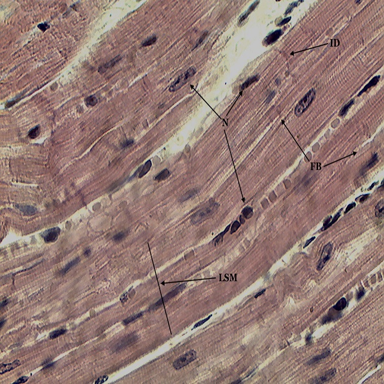
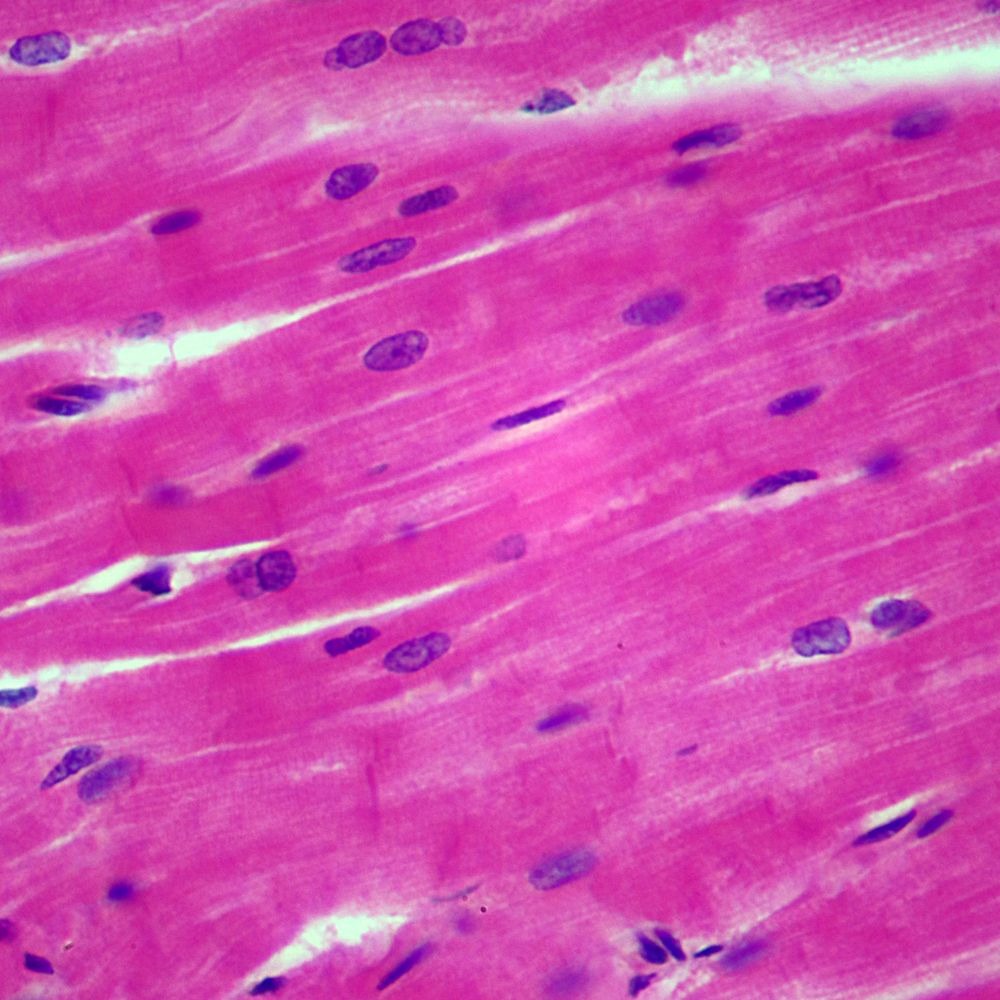
Each cardiomyocyte contains one or more nuclei situated centrally. It’s a hub from where cellular activities get directed. Surrounding the nucleus, the cytoplasm is abundant with mitochondria. These are the cell’s powerhouses, supplying energy for constant contractions.
Another standout feature is the presence of striations. These are alternating light and dark bands visible under the microscope. They reflect the aligned myofilaments within the cells, essential for muscle contraction.
Most distinctively, cardiac muscle cells have intercalated discs. These special junctions link cells together, ensuring a unified contraction across the heart muscle. They also facilitate electrical signals’ swift passage. It makes the heart beat in rhythm.
The combination of these traits equips cardiac muscles with extraordinary endurance. They keep the heart pumping efficiently, without succumbing to fatigue swiftly. Through microscopic investigation, the unique organization and resilience of cardiomyocytes become evident, highlighting their critical role in sustaining life.
The Role of Cardiac Muscles in the Human Body
The heart’s engine, cardiac muscle, plays an essential role in our well-being. The cells work together, contracting to pump blood throughout the entire body. This flow is vital for delivering oxygen and nutrients to other tissues. Without the cardiac muscle’s pumping action, organs would starve, and waste would build up.
Cardiac muscle’s role goes beyond basic pumping. It maintains blood pressure and ensures a steady heartbeat. Every moment, heart muscles adapt to our body’s demands. When we exercise, they contract faster and harder, increasing blood flow to muscles needing more oxygen. During rest, the contractions are softer and steady, conserving energy.
Even subtle changes in our emotional state cause shifts in cardiac activity. The muscle responds to adrenaline by increasing heartbeat, preparing us for ‘fight or flight’. But it also can be damaged; lifestyle, diet, and stress can affect its health.
Keeping the cardiac muscle healthy is crucial. Regular exercise, a balanced diet, and monitoring health are key. We need to understand its central role to take better care of our heart. The cardiac muscle under the microscope reveals not just an organ but a symbol of life itself.
Examining Cardiac Muscle Under the Microscope
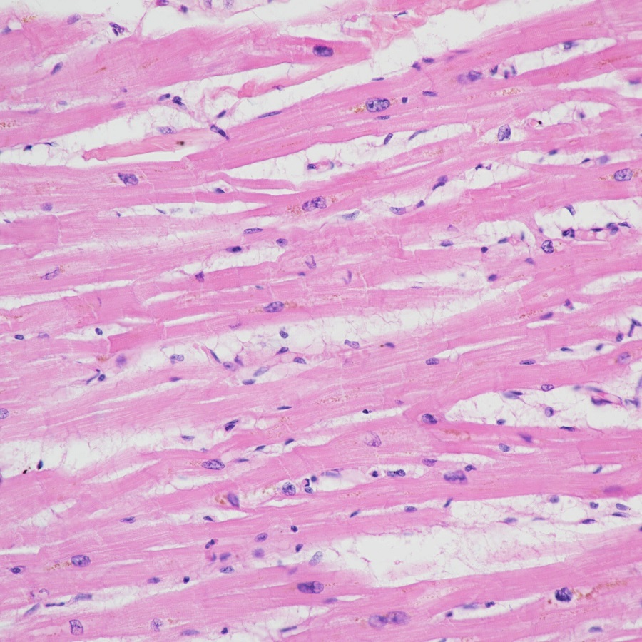
When we place cardiac muscle under the microscope, its true complexity unfolds. This examination invites us into a world where form meets function in stunning detail. Observing cardiac muscle under microscope reveals an intricate architecture. This design is vital for the heart’s relentless workload.
Each cardiomyocyte emerges as a masterpiece of biological engineering. Under magnification, their branch-like structures come into sharp relief. These branches form seamless connections with fellow cells. It’s a configuration that enables the heart’s synchronous beating.
Striated patterns crisscross the cells, showing the alignment of myofilaments. These are the tiny engines that drive contraction. Striations are the signatures of a healthy, functioning muscle. They indicate precision in the muscle’s micro-universe.
The most fascinating aspect is perhaps the intercalated discs. Only seen under high-power lenses, these junctions embody the heart’s electrical sync. They are like circuit connectors, ensuring rapid signal transmission. This lets each heartbeat maintain a steady, life-giving rhythm.
By observing cardiac muscle under microscope, we gain insights into its endurance. The high density of mitochondria is striking and tells a story of energy and resilience. Each cell is a powerhouse, drawing on these organelles to sustain non-stop work.
In conclusion, a microscopic view of cardiac muscle uncovers a tapestry of cellular interaction. It teaches us about the muscle’s capacity to endure and perform. It is not just a glimpse into the heart but an exploration of life sustaining machinery at work.

Structural Comparison: Cardiac Muscle vs. Skeletal Muscle
When examining cardiac muscle under microscope, we see its uniqueness. But how does it differ from skeletal muscle, which moves our bones? Both are muscle tissues, yet they serve distinct roles.
Cardiac muscle, found only in the heart, beats without rest. It pumps blood with a rhythm, adjusted by our body’s needs. Its cells are short, branched, and tightly connected. This design lets the heart beat as one powerful unit.
Skeletal muscle, on the other hand, helps us move. It is attached to bones by tendons. Skeletal muscle cells are long, cylindrical, and not branched. They form bundles that pull on bones, creating movement.
Under microscope, striations show in both. Yet their patterns differ, hinting at functions they serve. In cardiac muscle, the striations have a unique order, essential for nonstop work.
One more difference is visible in the cells. Cardiac muscle cells have one or more nuclei located at the center. Skeletal muscle fibers generally have many nuclei, spread along the edges.
Another distinct feature is intercalated discs, only in cardiac muscle. They do not appear in skeletal muscle. These discs allow signals that control heartbeats to pass quickly.
In summary, though both are muscles, their structure is suited to their jobs. Cardiac muscle, under microscope, reveals a network built for endurance and constant activity. Skeletal muscle fibers demonstrate a structure for controlled and forceful movements.
The Microscopic World of Intercalated Discs
Intercalated discs are crucial in the cardiac muscle’s microscopic landscape. These specialized structures are visible when we examine cardiac muscle under microscope. They appear as dark connecting lines between cardiomyocytes. Their role? To bind heart cells firmly, ensuring a tight, secure connection among them.
Under high magnification, the discs’ complex features stand out. They house gap junctions and desmosomes. Gap junctions let electrical impulses travel swiftly between cells. This ensures the heart beats in a coordinated fashion. Desmosomes, on the other hand, provide structural support. They keep cardiomyocytes attached during the intense work of beating.
Intercalated discs also help the heart react as a whole. When one cell contracts, the discs transmit the force to neighbor cells. This domino effect results in a synchronized contraction of the heart muscle.
Observing these discs under a microscope reveals the heart’s capacity for teamwork. Each cell is an individual player, yet all work in unison thanks to intercalated discs. It’s a microscopic view into the togetherness that powers every heartbeat.
In essence, intercalated discs are the cornerstone of the cardiac muscle’s reliable rhythm. They exemplify teamwork at the cellular level, essential for our survival.
Cardiac Muscle Adaptations and Endurance
Cardiac muscle responds to life’s demands with impressive adaptations. When under a microscope, we catch glimpses of these changes. These adaptations grant the heart incredible endurance. It allows it to work tirelessly, pumping blood constantly throughout our lives. Observing cardiac muscle under a microscope, we can appreciate these adjustments more deeply.
One key adaptation is the high number of mitochondria. These tiny organelles produce energy cells need for contraction. In cardiac cells, their abundance ensures ample energy supply for non-stop activity. Under the microscope, we see them densely packed, ready to fuel the muscles.
Another notable adaptation is hypertrophy, or cell enlargement. This occurs when the heart muscle strengthens in response to increased workload, such as during exercise. Under the microscope, these larger cells show just how the heart adapts and grows stronger over time.
Cardiac muscles also have a remarkable repair ability. While they cannot regenerate like some tissues, their cells can adapt to stress or injury. They do so by reorganizing their internal structure, which we can see as changes under a microscope.
Finally, the presence of a vast network of capillaries ensures that the muscle receives a constant supply of oxygen and nutrients. Microscopic examination reveals this intricate web, highlighting how well the cardiac muscle is supported.
In conclusion, the endurance of cardiac muscle is not by chance. It’s a result of constant adaptation at the microscopic level. This adaptation ensures our heart beats strong, day and night, throughout our lives.

The Impact of Diseases on Cardiac Muscle Microstructure
Cardiac muscle, viewed under the microscope, unveils a finely tuned structure. Yet diseases can disrupt this delicate balance. Chronic conditions modify the microscopic appearance of cardiac muscle. Understanding these changes aids in grasping the disease’s impact. It also helps in recognizing the signs early.
Diseases like cardiomyopathy alter the muscle’s texture, seen clearly under a microscope. The once orderly striations become erratic. Muscle fibers may appear swollen or fragmented. These are signs of the heart struggling to function.
Another example is atherosclerosis. It can reduce blood flow, starving cardiac muscle cells of oxygen. Under high magnification, this damage is visible. Cells may seem shrunken or have less defined borders. These conditions signal the suffering of tissues.
In cases of myocardial infarction, commonly known as a heart attack, dramatic changes occur. Observing cardiac muscle under microscope post-attack reveals dead zones. Such areas lack the uniform appearance of healthy heart tissue.
All these disease-induced alterations lessen cardiac muscle’s ability to pump blood. Recognizing these microscopic signs helps in diagnosing and treating heart disease. Regular check-ups can spot these signs early, leading to better outcomes.
In conclusion, diseases wreak havoc on the microstructure of cardiac muscles. Microscopic observation serves as a powerful diagnostic tool. It allows us to see the effects of disease up close. Knowledge of these changes empowers us to understand and combat cardiac diseases better.