Introduction to Microscopy
The study of cells under microscope starts with microscopy. This is the science of using microscopes to view tiny structures. The term ‘microscopy’ means ‘to look at the small.’ It allows us to explore worlds too small for the naked eye. We can see the intricate details of cells. This is crucial for medical research, biology, and education.
Microscopes come in many forms. There are simple ones and complex ones with high tech features. They make the invisible visible. We magnify the size of cells many times over. We peer into the building blocks of life itself.
When we see cells under microscope, we learn about their function and structure. This helps us understand how life works on a basic level. Whether for a student or scientist, microscopy is a gateway into the microscopic world. It empowers us to discover and to invent.
Types of Microscopes Used to View Cells
To view cells under microscope, we use different types of microscopes. Each kind has unique features and applications. The common ones include light microscopes, electron microscopes, and fluorescence microscopes.
Light microscopes are the most widely used. They magnify cells using visible light. You find these in high school labs and medical offices. Beginners start with these as they are user-friendly. Students see the basic structure of cells with light microscopes.
Electron microscopes go deeper. They use beams of electrons for higher magnification. With them, you see cell details invisible to light microscopes. Electron microscopes come in two main types: transmission and scanning. Transmission electron microscopes (TEM) give detailed cell interior views. Scanning electron microscopes (SEM) offer 3D images of cell surfaces.
For live cells and their activities, we use fluorescence microscopes. They use special dyes that glow under specific light. This lets us watch live cells and their processes in real-time. Researchers understand cell behavior better as a result.
Compound microscopes are also worth mentioning. They have multiple lenses. This increases their magnification power. Typically, they serve well in advanced educational settings.
In summary, the choice of microscope depends on the need. Whether it’s a simple look at cell structure or a deep dive into cellular activities, there’s a microscope suited for the task. With these tools, we expand our view of the small and uncover the mysteries of cells under microscope.

Preparing Cells for Microscopic Examination
Before examining cells under a microscope, one must prepare them appropriately. This crucial step ensures the cells are visible and distinguishable. Here are the key steps in the cell preparation process for microscopic examination:
- Collection: First, gather the cells. For human cells, this may involve a cheek swab. For plant cells, a leaf section might suffice.
- Fixation: Cells must remain in place. Fixing them to a slide helps preserve their structure during viewing.
- Permeabilization: For certain stains to enter, we sometimes need to make the cell membranes more porous.
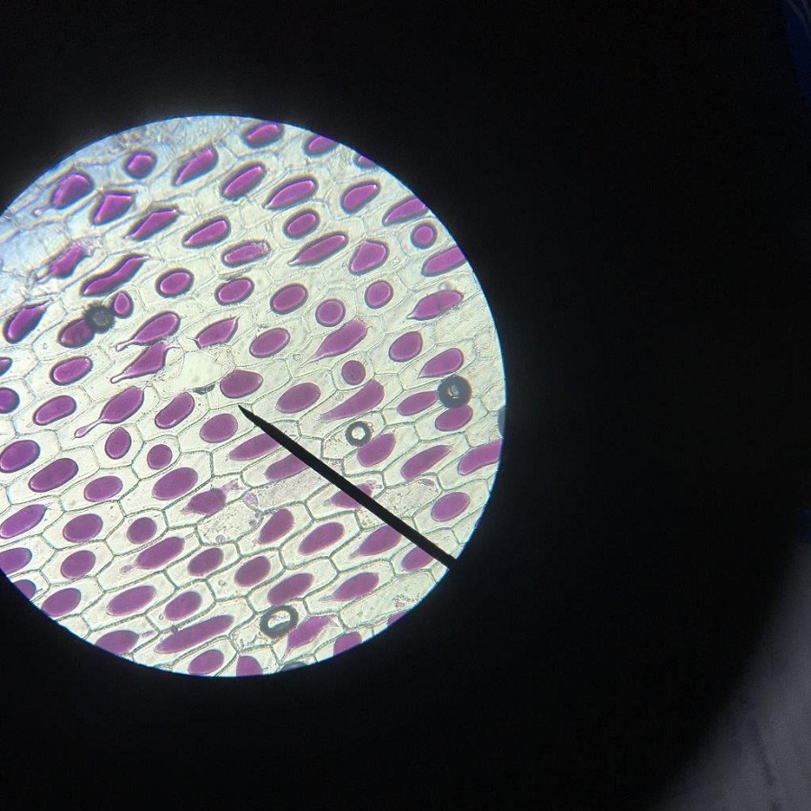
- Staining: Stains and dyes add contrast. They highlight different parts of the cell. This makes structures like the nucleus stand out under the microscope.
- Mounting: Finally, cover the sample with a cover slip. This protects the cells and keeps them flat for optimal viewing.
Each step needs care, as poor preparation can mask or distort features. With cells prepped well, we can then place them under microscope to observe their marvels.
The Human Cell Structure Observed Under Microscope
The human cell is complex and full of minute details, a true marvel when observed under a microscope. By placing cells under a microscope, we get to explore the cell structure in great detail. This includes the outer membrane, cytoplasm, nucleus, and various organelles. Each plays a vital role in the cell’s overall function.
The cell membrane encloses the cell’s contents and controls what enters and exits. Through the microscope, it appears as a distinct outline, enclosing the mysteries within. The cytoplasm, a jelly-like substance, fills the cell and supports the organelles. Here, we see the hustle of cell life with elements moving around.
The nucleus, usually the most prominent feature, houses DNA and manages cell activities. Under the microscope, it often stands out because of its size and shape. Mitochondria, known as the powerhouses, are also visible. They look like tiny beans working tirelessly to produce energy for the cell.
As for the endoplasmic reticulum, it appears as a network, crucial for making and shipping proteins and lipids. A close-up on these can be fascinating, showing the paths along which the cell’s products move. The Golgi apparatus, resembling a stack of pancakes, packages proteins for transport.
We can even see lysosomes and peroxisomes—tiny recyclers breaking down waste. They can sometimes be seen as small dots or bubbles. When we see these structures, every dot and thread reveals how cells function and maintain life.
Human cells under microscope give us insight into diseases, too. Abnormalities become apparent, like irregular nuclei in cancer cells. This is why microscopy is so vital in medical diagnostics and research. It lays the groundwork for understanding health and developing treatments.
In summary, viewing human cells under microscope offers an extraordinary glimpse into our own biology. It’s a journey into the inner workings of life at a scale invisible to the naked eye, yet pivotal to our existence. The intricate dance of cellular life, witnessed at such a close range, never ceases to amaze.
Observing Plant Cells Through the Microscope
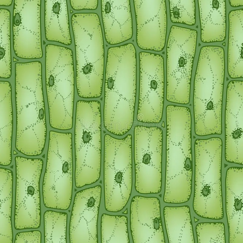
When observing plant cells under microscope, we unveil a world of rigid structures and vibrant green chloroplasts. These cells, with their cell walls made of cellulose, appear different from the flexible membranes of animal cells. Here is what we typically notice when peering into the microscopic world of plants:
- Cell Wall: Plant cells have a sturdy outer layer providing shape and support.
- Chloroplasts: These green structures harbor chlorophyll, enabling photosynthesis to occur.
- Central Vacuole: A large, fluid-filled sac that stores nutrients and helps in maintaining cell turgor.
- Plasmodesmata: Tiny channels traversing cell walls, allowing communication and transport between cells.
The rigidity of the cell wall and the presence of chloroplasts are standout features. The microscope reveals the grid-like pattern of the cell wall and the dynamic green chloroplasts moving within the cell. The large central vacuole, often dominating the cell’s interior, is easily visible. It becomes apparent that plant cells are well-equipped for their role in photosynthesis and structural integrity.
Looking at plant cells under microscope, we also witness stomata, the small pores on leaves for gas exchange. These tiny openings are critical for the plant’s breathing and transpiration processes.
To sum up, using a microscope to observe plant cells provides a fascinating view into the structures that support plant life. It sheds light on how plants absorb sunlight, make food, nourish themselves, and interact with their surroundings. This microscopic examination is essential for botanists and horticulturists as they study plant health and function.

Microscopic Examination of Microorganisms
Just as with plant and human cells, observing microorganisms under the microscope opens up a new world. Microorganisms are incredibly diverse, ranging from bacteria and viruses to fungi and protozoa. Here’s what we typically uncover when studying these tiny life forms:
- Bacteria: With a microscope, we see bacteria’s simple structures and various shapes. These include spheres, rods, and spirals.
- Viruses: Much smaller than bacteria, viruses often need electron microscopes. This reveals their complex geometric shapes.
- Fungi: We notice fungi’s thread-like structures through a microscope. They can display an array of intricate networks.
- Protozoa: In water drops, we find protozoa. We notice their movement and feeding methods.
Microscopes help us identify and classify microorganisms. They let us see how these organisms interact with their environment. They also aid in understanding diseases and their treatments.
Medical professionals rely on microscopy to identify pathogens in samples. This is crucial for diagnosing infections. Microscopes also play a key role in microbiology research. They help to discover new microorganisms and study their behavior.
In essence, the microscopic examination of microorganisms is a key tool. It helps us to understand and combat diseases, and to unlock the secrets of the tiniest forms of life.
Photomicrography: Capturing Cell Images
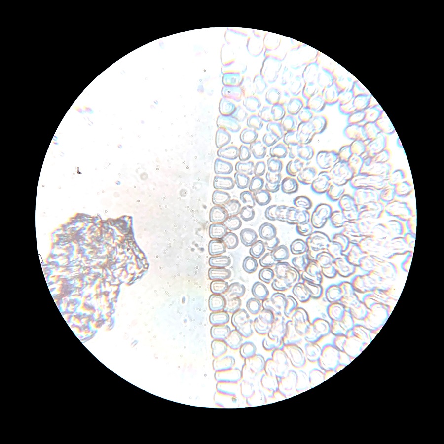
Photomicrography is the art of taking photographs through a microscope. This technique captures the stunning details of cells under microscope. It lets us share and analyze the tiny world of cells. Experts use special cameras attached to microscopes for this purpose.
Photomicrography requires skill and patience. You must adjust the lighting and focus carefully. Clear images depend on proper sample preparation and microscope settings. High-quality lenses and stable platforms are also vital.
Once the cells are photographed, software helps enhance the images. This brings out more detail in the structures observed. Researchers and educators use these images to study and teach about cell life. These photos also often appear in textbooks and scientific papers. They are vital for visual documentation in research.
Even amateur scientists can try photomicrography. Many microscopes now come with built-in cameras. This makes capturing cell images more accessible to hobbyists.
In conclusion, photomicrography turns cells seen under microscope into lasting images. These images inspire curiosity and lead to advances in science and medicine. They give us a visual record of the beauty and complexity of cells.
Understanding Stains and Dyes in Cell Visualization
When we place cells under microscope, stains and dyes are critical. They give color to parts of the cell that are otherwise clear. Here’s what they do and why they’re important:
- Contrast Enhancement: Dyes add color to cell components. This contrast helps us see details we’d otherwise miss.
- Specificity: Some stains bind to specific cell parts. For example, DNA dyes highlight the nucleus, making it stand out.
- Cell Differentiation: Different stains can show us separate parts of a cell. They can dye the nucleus one color and the cytoplasm another. This way, it becomes easier to tell them apart.
- Visualization of Cell Processes: With fluorescent dyes, we can watch cells live. This lets us observe how they move and interact.
- Medical Diagnosis: Stains make abnormal cells more noticeable. This is key in identifying diseases under a microscope.
In the lab, biologists use a variety of dyes. Some common ones include crystal violet for bacteria and iodine for starch in plant cells. Others, like hematoxylin and eosin (H&E), are standard for human tissue. Fluorescent dyes, like DAPI, bind to DNA and glow when lit up.
Each stain and dye works differently. Some need to fix the cells first. Others can enter living cells without harm. The choice depends on what parts of the cell we need to see. The right stain can turn a fuzzy view into a clear picture of the cell’s life.
In essence, understanding how stains and dyes work enhances our microscopic exploration. They allow us to see the tiny details that tell the big story of life at a cellular level. This understanding is vital for research and education in biology and medicine.

Exploring Cell Movement and Behavior under the Microscope
When we view cells under microscope, we don’t just see static images. We observe how cells act and interact. This reveals life’s dynamics in fascinating detail. Here’s how cell movement and behavior become a visual feast under the microscope:
- Cell Motility: This term describes how cells move. Some, like white blood cells, crawl to fight infections. Watching these cells move is like seeing an army in action.
- Chemotaxis: Cells often move toward or away from chemicals. This is chemotaxis. Under the microscope, we watch cells navigate like tiny ships, sailing towards nutrients or away from toxins.
- Cell Division: Microscopes let us view cell division live. We watch one cell split into two. It’s like witnessing the moment of birth over and over.
- Cell Interaction: Cells don’t live alone. We see them touch, stick, and talk to each other through signals. This social side of cells is key for understanding how tissues and organs form.
- Behavior Response: Cells react to their environment. They respond to light, temperature, and touch. Under high-powered lenses, we see these reactions in real time. It’s like watching cells make decisions.
Watching cells move and interact teaches us about life at its most basic level. It helps scientists discover how diseases spread and heal. It also reveals how groups of cells work together to keep organisms alive and healthy.
In summary, studying cell movement and behavior under the microscope is essential. It helps us unravel the mysteries of cellular life. With this knowledge, we push the boundaries of science and medicine further.
Conclusion and the Future of Cellular Microscopy
The journey we’ve taken peering at cells under microscope reveals the unseen. We’ve explored how microscopes unlock the tiny world of cells. We’ve prepared cells, observed their complex structures, and captured images. Along the way, we’ve seen the importance of stains and dyes. We’ve watched cells come alive, moving and behaving in ways that tell the story of life itself.
Looking ahead, the future of cellular microscopy is bright. Advancements in technology promise even deeper insights. Novel microscopes will show us more detail, faster and in 3D. We’ll witness cell interactions in ways we can’t yet imagine. These tools will power discoveries in medicine, biology, and beyond.
Stains and imaging tech will evolve. They will make cell parts stand out like never before. We’ll track live cells over time, watching diseases form and heal. This couldn’t be more important for developing new treatments.
As we push the boundaries of what we can see and understand, one thing is clear. The microscopic examination of cells will continue to be a cornerstone. It will guide us in understanding the complex dance of life. Learning more about our world, improving health, and curing diseases will follow. The magic we discover with cells under microscope is just the beginning.
