Introduction to Compound Light Microscopes
A compound light microscope is a popular tool in science labs. It helps us view very small objects in detail. This microscope uses light to illuminate samples. It has several labeled parts that work together to magnify objects. Students and researchers use it to study cells, microorganisms, and other tiny structures
The compound light microscope has two sets of lenses: the ocular and objective lenses. The ocular, or eyepiece, is where you look through. The objective lenses are on a revolving nosepiece. They come in different powers of magnification. The stage holds the sample, and stage clips keep it in place. Below the stage is the condenser, which focuses light onto the sample. The illuminator is the light source of the microscope.
Using microscope starts with placing the sample on the stage. Next, you select the right objective lens. You then adjust the focus using two knobs. The coarse adjustment knob makes large changes. The fine adjustment knob makes small, precise changes. Proper focus is key to clear, detailed images.
To keep a compound light microscope working well, handle it with care. Keep the lenses clean and store the microscope in a safe place. This ensures it stays accurate and lasts a long time.
In the following sections, we’ll dive deeper into the parts of a compound light microscope. We will explore how each component contributes to the device’s function. We’ll also cover maintenance tips and compare this microscope to other types.
Core Components of a Compound Light Microscope
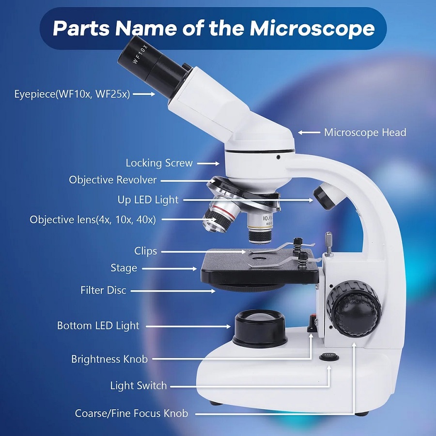
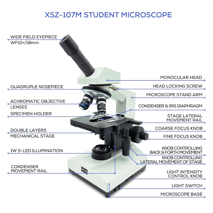
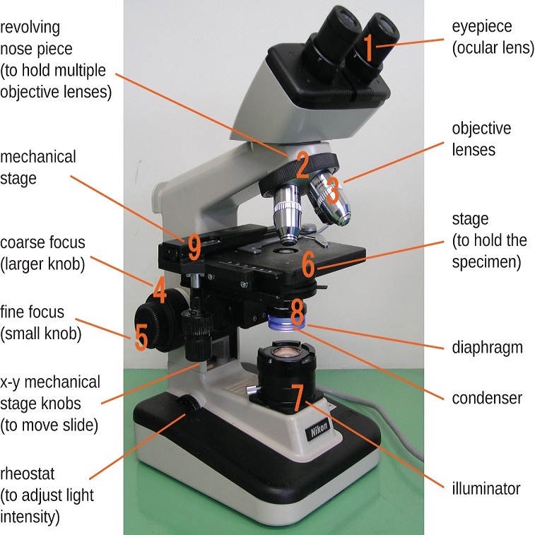
Compound light microscopes are intricate scientific tools. They comprise several core components that work in unity to enhance the viewing of tiny objects. The main parts of a compound light microscope include the eyepiece or ocular lens, objective lenses, stage, stage clips, condenser, and illuminator. Each of these components plays a critical role in the microscope’s functioning. Let’s go over these labeled parts and understand their significance.
Eyepiece or Ocular Lens
The eyepiece is where users place their eyes to view the magnified image. It typically contains one or more lenses that further magnify the image projected by the objective lenses. The eyepiece’s magnification is usually 10x.
Objective Lenses
Underneath the eyepiece sits a rotating nosepiece with various objective lenses. Each lens provides a different level of magnification, such as 4x, 10x, 40x, or 100x. Scientists select the lens based on their required magnification.
Stage and Stage Clips
Samples sit on the stage, which is a flat platform. Stage clips secure the samples in place during observation. The stage’s position can be adjusted to bring the sample into the objective lens’s focus range.
Condenser
Situated below the stage, the condenser gathers and focuses light from the illuminator onto the sample. This part helps in improving the clarity and contrast of the viewed specimen.
Illuminator
The illuminator acts as the light source for the microscope. It shines light through the sample, making the tiny details visible through the lenses. The brightness of the illuminator can often be adjusted, depending on the sample’s transparency and the magnification used.
Understanding each component’s function enhances the operator’s ability to utilize the microscope effectively. These parts are deliberately designed to work together to give researchers and students a closer look at the smaller wonders of our world. In the next sections, we will delve into the focusing mechanisms and the optical system that further define the capabilities of a compound light microscope.
Focusing Mechanisms
A compound light microscope has two main focusing mechanisms. These are the coarse adjustment knob and the fine adjustment knob. Both are crucial for achieving a clear, sharp image.
Coarse Adjustment Knob
The coarse adjustment knob is the larger of the two knobs. It raises or lowers the stage quickly. This brings the sample closer to or further from the objective lenses. It’s best used when you begin observing your sample. You first use this knob to get a rough focus.
Fine Adjustment Knob
The fine adjustment knob is smaller and allows for more precise movements. It fine-tunes the focus after you’ve used the coarse adjustment knob. This knob adjusts the stage or lenses very slightly. It sharpens the image for the best clarity. You use this knob to make the image as clear as possible.
Using both knobs together helps you see the details of the specimen. It’s important to start with the coarse knob. Then, once you have a general focus, switch to the fine adjustment knob for a detailed view. These mechanisms work together to reveal the tiny wonders under the lens of the compound light microscope.
Optical System and Magnification
The optical system of a compound light microscope defines its magnification abilities. It involves the eyepiece and objective lenses. Together, these lenses magnify the sample image several times.
Dioptric Adjustment
The dioptric adjustment aligns the focus between both eyes. It accounts for the difference in vision that a user might have. This feature is often part of the eyepiece. It ensures a crisp image for each viewer’s individual needs.
Magnification Range
A compound light microscope’s magnification range can be extensive. It typically starts at 40x. It can go up to 1000x or more. To achieve this, users combine the eyepiece magnification with the chosen objective lens. The total magnification is the product of these two. For instance, a 10x eyepiece and a 40x objective lens give 400x total magnification. Adjusting these components allows for various levels of detail to be examined.
Maintenance and Proper Use
Proper maintenance and usage are essential for the longevity and accuracy of a compound light microscope. The following points explain how to keep this crucial lab instrument in top condition.
Cleaning Lenses and Components
Lenses capture and magnify the minute details of samples. They require careful cleaning. Use a soft, lint-free cloth for wiping. Gently remove any dust, oil, or fingerprints. Do not use harsh chemicals; they might damage the lens coatings. Cleaning solutions designed for optical components are best. For other parts of the microscope, such as the stage and body, use a mild detergent and a damp cloth.
Handling and Storage Best Practices
Handle the microscope with great care. Always carry it with both hands — one on the arm and the other supporting the base. Keep the device covered when not in use. This protects it from dust. Store it in a dry, temperature-controlled environment. Make sure there’s no direct sunlight as it can damage the optical parts. Regular checks are necessary. They ensure all components function well and are properly aligned.
In summary, proper handling, cleaning, and storage are key to the microscope’s performance and service life. This care allows researchers and students to conduct their studies without interruptions caused by equipment issues. These best practices ensure that the compound light microscope labeled remains a reliable tool in scientific investigation.
Comparison with Other Types of Microscopes
Understanding the capabilities of a compound light microscope labeled is helpful. Yet, it’s equally important to see how it compares to other types. Two common alternatives are stereo microscopes and electron microscopes.
Stereo Microscopes
Stereo microscopes, known as dissecting microscopes, have two eyepieces. They provide a 3D view of the specimen. Unlike compound microscopes, they offer lower magnification. This is usually up to 40x. They are ideal for viewing larger, solid objects. Such as insects, plants, and circuit boards. Their design allows for working space between the object and the lens. This space makes it possible to perform tasks. Like dissection, under the microscope.
Electron Microscopes
Electron microscopes use beams of electrons instead of light to create an image. They have much higher magnification and resolution. Some can magnify objects up to two million times. They let us see tiny details. Even individual atoms can be observed. But, electron microscopes are much larger and costlier than light microscopes. They require special environments and trained operators. Because of this, they’re mainly found in advanced research facilities.
Knowing the differences between microscope types aids in choosing the right one for a study. Each type serves a specific purpose. This guide undershadows the importance of a compound light microscope labeled in the scientific community. It also highlights options for various research needs.
Advancements in Microscopy
The field of microscopy has seen remarkable advancements, enhancing our ability to study the microscopic world. Continuous innovation has led to more sophisticated compound light microscopes labeled with features that extend their use and capabilities. Let us highlight some key developments in this area.
Digital Integration
Today’s microscopes often come with digital integration. This technology allows users to connect microscopes directly to computers. It enables them to capture images quickly and store them easily. With digital cameras and software, capturing high-resolution images is straightforward. Live streaming of microscopic images for educational purposes has also become common.
Enhanced Imaging Capabilities
Imaging quality has greatly improved with the advent of higher-resolution cameras and advanced lighting techniques. Enhanced imaging capabilities include better contrast methods, such as phase contrast and differential interference contrast, which offer a deeper view of samples. Fluorescence microscopy has also evolved. It uses specific light to illuminate biological markers in samples. These improvements support complex research and detailed analysis of specimens.
The leaps in microscopy technology have significantly contributed to scientific progress. Having a compound light microscope labeled with these cutting-edge features ensures precise and efficient observations. This advancement opens new doors for exploration at scales that were once unfathomable.