Understanding the intricate layers of our skin has always fascinated scientists, dermatologists, and beauty enthusiasts alike. By examining skin under microscope, we gain invaluable insights into its structure, health, and the effects of various treatments. Whether you’re curious about how your skincare products work or interested in the science behind skin conditions, exploring skin under a microscope can reveal truths that are invisible to the naked eye. In this comprehensive guide, we’ll delve into the fascinating world of skin microscopy, uncovering its benefits, techniques, and applications in both medical and cosmetic fields.
 The Basics of Skin Structure
Our skin is an engineering marvel and serves many roles, from protecting internal organs to regulating body temperature. To understand what “skin under microscope” reveals, let’s start with the basics. Skin is the body’s largest organ and has a complex structure composed of three primary layers: the epidermis, dermis, and hypodermis.
Epidermis
The outermost layer, the epidermis, forms the barrier against environmental threats. Under a microscope, it shows a tight grid of cells renewing constantly. These cells, known as keratinocytes, move from the bottom to the top of the epidermis, creating a tough outer surface.
Dermis
Below the epidermis lies the dermis. This middle layer houses blood vessels, nerves, hair follicles, and sweat glands. Collagen and elastin fibers, providing skin its elasticity and strength, are visible here under higher magnification.
Hypodermis
The deepest layer, the hypodermis, contains fat and connective tissues that cushion and insulate the body. It’s less dense under the microscope, showing larger, spaced-apart cells that store energy and retain heat.
By examining the “skin under microscope”, professionals can learn a lot about a person’s health and skin conditions. Microscopic analysis helps to understand how healthy skin functions and what goes wrong during skin diseases.
A Closer Look at Skin Cells under the Microscope
When examining skin under a microscope, the complexity of skin cells becomes apparent. Additional insights can be drawn by focusing on the intricate details of these cells.
Visualizing Keratinocytes
Under high magnification, keratinocytes reveal their life cycle. You can witness new cells forming and older ones flaking off. This process is constant and contributes to our skin’s regeneration.
Examining Melanocytes
Another cell type visible under the microscope is the melanocyte. These cells produce melanin, which determines skin color and offers protection against UV rays. They appear as darker spots amidst keratinocytes.
Observing Langerhans Cells
Langerhans cells are part of the immune system and key under the microscopic lens. These cells detect and fight off foreign invaders, such as bacteria and viruses.
Identifying Fibroblasts in the Dermis
Moving deeper, in the dermis, fibroblasts produce collagen and elastin. These fibers give skin its strength and elasticity and look like threads woven together under the microscope.
By closely looking at skin cells under the microscope, one can appreciate the dynamic and protective nature of skin. Each cell type has a specific role that maintains the skin’s health and vitality. Understanding these roles is crucial in recognizing changes that may indicate skin conditions or diseases.
 Understanding the Different Layers of Skin
To appreciate the depth of what we can see under a microscope, consider the distinct layers of our skin. Each one, from surface to base, has unique cells and functions that contribute to our overall health. Let’s dive deeper into this intricate world.
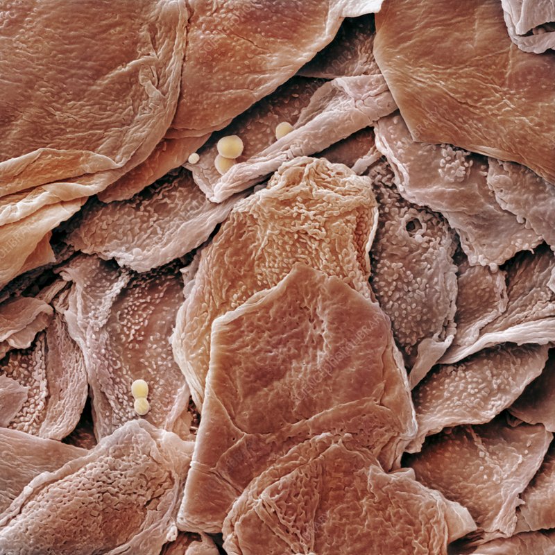
Epidermis: The Protective Barrier
This topmost layer offers the first line of defense against the environment. It boasts a protective battalion of keratinocytes. These cells are born deep within the epidermis and travel upward, forging a shield by the time they reach the surface. Dead and flattened, they flake away, making room for new guardians.
Dermis: The Supportive Network
Under the epidermis lies a more complex story. The dermis provides structural support with its network of collagen and elastin fibers. It hosts the machinery for senses and cooling; nerves and sweat glands make their home here. Its blood vessels nurture the epidermis above, ensuring vitality.
Hypodermis: The Insulating Layer
A deeper dive brings us to the hypodermis, a softer layer made primarily of adipose tissue – our natural insulation and shock absorber. It’s where energy reserves are stored and where body heat is retained, seen microscopically as more spaced-apart cells.
By understanding these layers, we grasp how ‘skin under microscope’ allows experts to detect anomalies that may point to skin conditions. Knowing each layer’s role clarifies why certain symptoms appear and provides clues for potential treatments. It’s like deciphering a code where each layer holds part of the secret to overall skin health.
Common Skin Conditions Through the Lens of a Microscope
When skin ailments occur, ‘skin under microscope’ becomes a powerful diagnostic tool. Microscopic examination can unveil underlying issues not visible to the naked eye.
Acne and Blocked Pores
For instance, acne, a prevalent skin condition, shows up microscopically as clogged pores. Oil, dead skin cells, and bacteria fill these pores, creating the well-known pimple or blackhead. Inspection under a microscope can help determine the severity and type of acne.
Eczema and Skin Flakes
Eczema, marked by itchy, inflamed skin, also takes on a unique appearance when magnified. The microscope can reveal excessive skin flaking and an irregular skin barrier that may contribute to the condition.
Psoriasis and Cell Turnover
Psoriasis, another common condition, is identified by rapid skin cell turnover. Under the microscope, thick, silvery scales become visible, indicating excess cell production and buildup.
Rosacea and Blood Vessels
Rosacea, which causes redness and visible blood vessels in the face, is another condition where microscopes are useful. They can expose the enlarged blood vessels that lead to the persistent redness seen in rosacea.
By observing ‘skin under microscope’, health professionals can learn much more about these conditions. This insight helps guide effective treatments and better understand each condition’s impact on skin health.
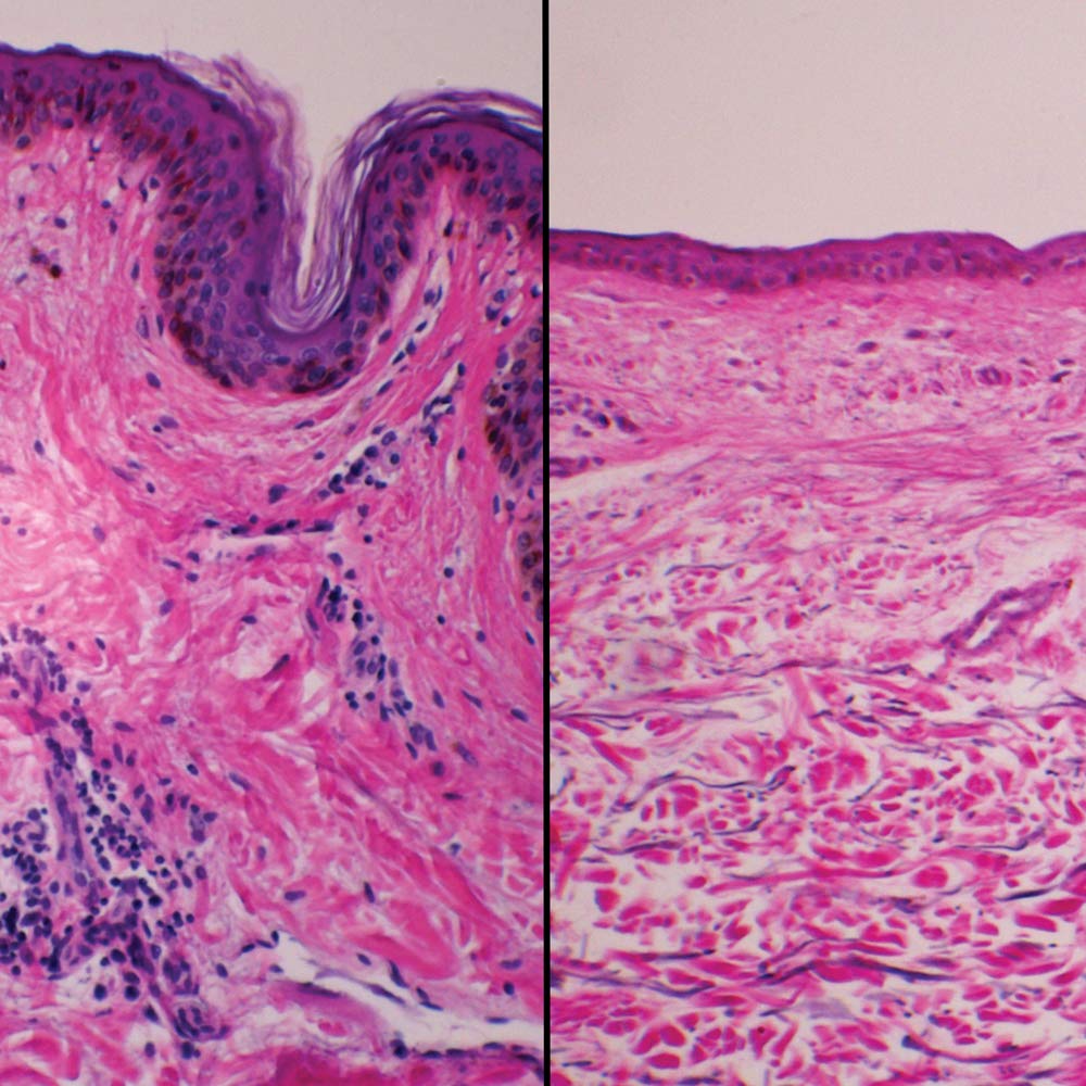
 The Impact of Aging on Skin Microstructure
Aging is a natural process that affects every organ, including our skin. A microscope reveals how aging changes skin’s microstructure significantly. Here are key effects:
Changes in the Epidermis
With age, the epidermis thins. Cell turnover slows, and fewer keratinocytes reach the surface. This makes skin appear more translucent.
Degradation of the Dermis
Collagen and elastin decrease in the dermis. This reduction in structural proteins makes skin less elastic and more prone to wrinkles.
Fat Redistribution in the Hypodermis
The hypodermis loses fat over time. This loss of volume can lead to sagging skin and a hollowed appearance.
Studying ‘skin under microscope’ shows us that aging impacts each skin layer. It affects the skin’s look, feel, and overall health. Recognizing these changes is vital for developing age-appropriate skin care routines.
How Diet and Lifestyle Affect Skin at the Microscopic Level
What we eat and how we live play a huge role in skin health. Beyond the visible, ‘skin under microscope’ tells an even more compelling story about the effects of diet and lifestyle.
The Role of Nutrition in Skin Cell Function
Nutrients fuel our skin cells. Under the microscope, one can spot the differences between well-nourished and poorly-nourished skin. A balanced diet rich in vitamins and antioxidants brings a healthy glow. It makes skin cells plump and vibrant, fighting off damage with ease.
A lack of healthy fats and hydration shows differently. Cells may look shrunken and function poorly. This causes the skin to lose elasticity and appear dull.
Lifestyle Choices and Skin Structure Integrity
Sleep, stress, and exercise impact skin structure. Good sleep helps cells repair and renew. This reflects in tight, orderly cell arrangements when viewed under high magnification.
High stress disrupts this order. It can weaken the skin’s barrier, making it prone to issues. On the other hand, regular exercise increases blood flow. This nourishes skin cells, keeping them healthy and resilient.
The Microscopic View of Skin Damage from UV Exposure
Ultraviolet (UV) rays from the sun or tanning beds do harm. Microscopes show this well. UV exposure can break down collagen and elastin. This causes premature aging and increases the risk of skin cancer.
Protection with sunscreen is visible under a microscope as a layer shielding the skin. Consistent use can preserve the skin’s underlying structure.
By looking at ‘skin under microscope’, it’s clear that daily choices shape our skin’s health. A good diet and lifestyle can maintain skin vitality and delay aging signs. Health experts use these microscopic clues to suggest better skin care practices.
Advances in Microscopy Techniques for Skin Analysis
As we delve into how ‘skin under microscope’ can provide valuable insights, it’s important to acknowledge the technological strides in microscopy. These advances have transformed our ability to analyze skin health and diagnose conditions with incredible precision.
Enhanced Image Resolution
Modern microscopes offer vastly improved image resolution. This clarity allows dermatologists to see cellular details that were once impossible to discern.
Digital Microscopy Integration
Digital integration means images can be stored and analyzed using software. This has streamlined the workflow for skin professionals, making the process more efficient.
3D Skin Imaging
Three-dimensional imaging techniques provide a comprehensive view. They reveal the skin’s topography from various angles, enhancing our understanding.
Live Cell Imaging
Technological progress has made it possible to observe live cells over time. Researchers can now watch how skin cells respond to treatments in real-time.
Confocal Microscopy
Confocal microscopy offers a deeper look into skin layers. By focusing on a single plane within the tissue, it provides a clearer picture of skin health.
These microscopy advancements offer a window into the microscopic world of skin health. They pave the way for more accurate diagnoses and tailored treatments. As the field grows, we can expect an even greater impact on our understanding of skin conditions.
Interpreting Microscopic Images of Skin for Better Diagnosis
The interpretation of ‘skin under microscope’ images is crucial for accurate diagnosis and treatment. With enhanced microscopy techniques, doctors can identify minute changes in skin cells that may indicate a range of conditions. Here’s how experts use microscopic images to improve diagnosis:
Spotting Abnormalities in Skin Cells
Under high resolution, abnormal cell shapes or sizes become clear. Doctors look for irregularities such as atypical pigmentation or unusual cell arrangements. These signs can signal skin disease or cancerous growths.
Recognizing Patterns of Inflammation
Patterns of cell clustering or disrupted layers often mean inflammation. This is typical of conditions like dermatitis or allergic reactions. The pattern gives clues about the cause and severity of the inflammation.
Tracing Blood Vessel Changes
Changes in blood vessels, like swelling or proliferation, are often seen with rosacea or other vascular disorders. The microscope helps in tracing these changes to assess the progress of the disease.
Assessing Collagen and Elastin Quality
The quality of collagen and elastin in the dermis could be affected by aging or sun damage. Microscopy lets doctors judge the damage extent and suggest suitable treatments.
Analyzing Glandular Structures
Microscopy can reveal if sweat or oil glands are blocked or if they have structural changes. This is key in diagnosing conditions like acne or seborrheic dermatitis.
Detecting Early Signs of Skin Aging
Spotting early signs of aging, such as thinning of the epidermis or fat loss in the hypodermis, can lead to preventive care. It helps in advising on proactive skincare routines.
The skillful interpretation of ‘skin under microscope’ images is critical for understanding complex skin conditions. It aids dermatologists and skincare professionals in crafting precise, effective treatment plans. The advancements in microscopic analysis are integral to the ongoing improvement of skin health diagnostics and therapies.
 Conclusion
Examining skin under microscope unlocks a world of detailed insights into one of our most vital organs. From diagnosing skin conditions and understanding the aging process to evaluating skincare products and enhancing medical research, the applications of skin microscopy are vast and impactful. By embracing the science behind what lies beneath your skin, you can make informed decisions about your skincare routine, seek timely medical interventions, and contribute to advancements in skin health and technology. Whether you’re a medical professional, a skincare enthusiast, or simply curious about the complex structures that protect and beautify us, exploring skin under a microscope offers a deeper appreciation of the incredible fabric that keeps us safe and glowing.