Understanding the intricate details of our blood is essential for diagnosing and monitoring various health conditions. By examining blood cells under microscope, medical professionals and researchers can gain invaluable insights into an individual’s health status. This detailed exploration delves into the types of blood cells, their appearance under a microscope, the techniques used for observation, and the significant applications of microscopic blood analysis. Whether you’re a student, healthcare professional, or simply curious about the microscopic facets of blood, this guide provides a thorough understanding of blood cells under microscope.
 A Brief Overview of Blood Cell Types
Before we dive deeper into the world of hematology and the role of microscopes, let’s first understand the types of blood cells. Blood cells are crucial for various body functions, and they fall into three primary categories: red blood cells (RBCs), white blood cells (WBCs), and platelets.
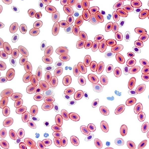
RBCs, also known as erythrocytes, are the most abundant cells in our blood. They carry oxygen from the lungs to the rest of the body and bring carbon dioxide back for exhalation. Under the microscope, RBCs appear as round, biconcave discs without a nucleus.
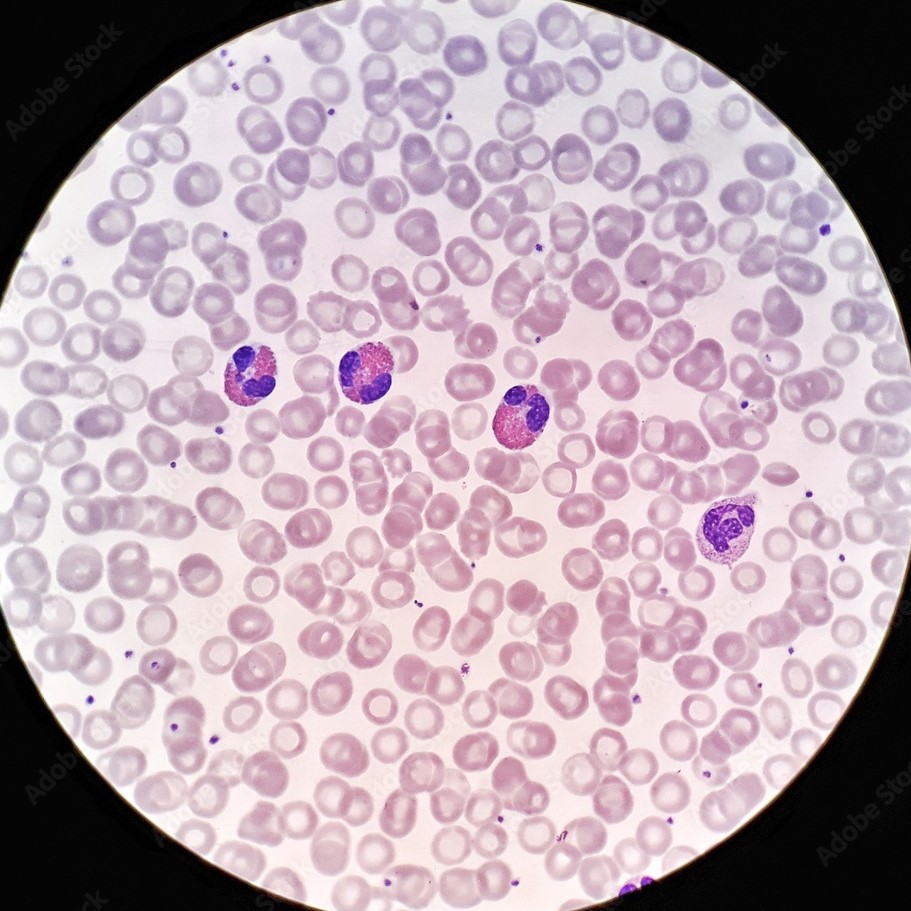
WBCs, or leukocytes, are the defenders of our body. They fight infections and play a vital role in the immune response. When examining blood cells under microscope, these cells are identifiable by their varied shapes and the presence of a nucleus.
Lastly, platelets, or thrombocytes, are tiny cell fragments. They play a critical role in blood clotting, a process vital to healing wounds. While observing blood cells under microscope, platelets can be seen as small, irregularly-shaped cells clustering together at injury sites.
In summary, each type of blood cell has a distinct role and appearance when viewed under a microscope. This overview sets the stage for a closer look at their microscopic features and highlights their importance in our overall health.
The Microscopic Structure of Red Blood Cells
The detailed examination of red blood cells under microscope offers a fascinating glimpse into their structure. They have a distinctive, biconcave shape with a diameter of about 6 to 8 micrometers. This unique shape increases their surface area, which is essential for efficient oxygen exchange. Red blood cells lack a nucleus, which allows more room for hemoglobin, the protein that carries oxygen.
Red blood cells exhibit extraordinary flexibility. This lets them navigate through the tiniest blood vessels, the capillaries. It’s under the microscope that we observe their remarkable ability to deform and pass through capillary beds barely larger than themselves. Their life span is around 120 days. After this, they are recycled by the spleen.
In the realm of hematology, studying red blood cells under microscope is key for diagnosing conditions like anemia. Anemia is characterized by a decreased number of red blood cells or insufficient hemoglobin. By evaluating the size, shape, and color of red blood cells under microscope, experts can uncover signs of various types of anemia and other disorders. This microscopic analysis is a cornerstone of medical diagnosis and monitoring.
 Identifying White Blood Cells and Their Functions
When we look at blood cells under microscope, white blood cells (WBCs) stand out. WBCs shield our body from infection. Each type has a unique role. Under the microscope, their various shapes and the presence of a nucleus make them distinct.
It’s fascinating to watch these cells in action. They patrol for foreign invaders like bacteria and viruses. Some WBCs engulf and destroy these intruders. Others create antibodies for long-term immunity. Their appearance can signal health issues. Experts look for abnormalities in size, shape, or number.
Microscopy helps diagnose infections, immune disorders, and blood cancers. This is due to the detailed visibility it provides. By identifying WBCs under microscope, doctors can take swift action to treat patients.
Platelets: The Clot Masters Under the Lens
When studying blood cells under microscope, platelets reveal their pivotal role in clotting. Tiny but mighty, these cell fragments are essential for stopping bleeding. They measure just 2-3 micrometers in diameter. Their small size belies their importance.
Under the microscope, platelets appear as small, irregular shapes. They swiftly cluster at injury sites. Here, they form a plug to prevent blood loss. Platelets also release chemicals crucial for healing and clot formation. Without these tiny actors, even minor wounds could cause serious issues.
Microscopy is vital in observing platelet function. It helps diagnose clotting disorders. These include conditions like hemophilia or thrombocytopenia. Both conditions can lead to excessive bleeding or clotting. By analyzing platelets under microscope, doctors can better assess and manage these disorders.
In summary, platelets play a critical role in our health. They are not just clot facilitators but also key to wound healing and blood stability. Microscopy brings their tiny world into view, aiding in crucial diagnoses and treatments.
The Staining Process in Blood Cell Microscopy
The art of blood cell analysis blossoms when staining techniques are applied in microscopy. Staining transforms the routine examination of blood cells under microscope into a vivid and insightful encounter. Let’s break down this critical process.
Stains are special dyes that adhere to different cell components, allowing them to stand out. During staining, a blood smear is treated with these dyes that give color to the cells and their parts. This enhancement is vital for observers. It makes the hidden features of blood cells visible and differentiates the cell types.
Typically, the staining procedure for blood cells includes the following steps:
- Preparation of the Blood Smear: Blood is spread thinly across a glass slide, creating a single layer of cells.
- Fixation: The smear is dried and fixed, often with methanol, to preserve the blood cells’ shape.
- Staining: Appropriate stains, such as Wright’s or Giemsa stain, are applied to the slide. These attach to different parts of the cells, like nuclei or membranes.
- Rinsing: Excess stain is gently washed off, leaving the helpful coloration behind.
- Observation: Once stained, the slide is ready for examination under the microscope.
This process not only aids in identification but also enhances the capacity to diagnose. Anomalies like size irregularities or structural abnormalities become easier to detect thanks to the contrasting hues. Conditions such as anemia, infections or blood disorders may reveal themselves through changes that staining highlights.
In short, staining brings a world of color to the study of blood cells under microscope, providing essential classification and diagnostic capabilities. For anyone analyzing blood cells, mastering staining is as critical as the observational skills needed to spot the subtlest of cellular nuances.
 The Role of Microscopy in Hematology Diagnosis
Microscopy stands as a cornerstone in hematology, the study of blood. It plays a crucial role in diagnosing various blood disorders. Viewing blood cells under a microscope gives healthcare professionals the ability to spot minute abnormalities that could indicate serious health conditions. Diagnostic hematology heavily relies on the detailed images microscopy provides.
Here are specific ways that microscopy aids in hematology diagnosis:
- Detection of Cell Abnormalities: Microscopy can reveal irregularities in size, shape, or structure of blood cells. These can be early signs of diseases such as anemia or leukemia.
- Quantifying Blood Cells: Accurate counts of red blood cells, white blood cells, and platelets are essential. Deviations from normal counts can be red flags for underlying issues.
- Identifying Parasitic Infections: Some parasites, like the ones causing malaria, can be identified when viewing blood cells under microscope.
- Assessing Blood Cell Health: The health of the cells can be assessed by their appearance. Against the enhanced contrast staining provides, the vitality of blood cells can be gauged.
- Supporting Treatment Decisions: Microscopic analysis informs treatment plans and tracks the effectiveness of therapies.
In short, microscopy is a vital tool in hematology. It uncovers hidden details that are often the key to diagnosis. Utilizing this tool, many blood-related health problems can be caught and treated at early stages. From spotting infections to tracking treatment response, the role of microscopy is indispensable in hematology diagnosis.
Advances in Microscopic Techniques for Blood Analysis
As experts in hematology harness the power of microscopes, they now encounter an array of advanced techniques. These advancements offer clearer, more detailed images of blood cells under microscope. A discussion of these techniques highlights the progress in blood analysis.
- High-Resolution Microscopy: This technique captures images with amazing clarity. It allows for close examination of blood cells, unveiling structures not seen before.
- Fluorescence Microscopy: In this method, cells are tagged with fluorescent markers. These bind to specific cell parts, lighting them up under the microscope’s special light.
- Confocal Microscopy: This creates sharp, 3D images of cells. It cuts through layers to focus on select parts, giving depth to blood cell analysis.
- Digital Imaging: Powerful software now aids microscopic study. It allows for enhanced image capture and analysis, not possible in the past.
The impact of these techniques is significant. They improve the detection and understanding of blood cell behavior. For instance, rare cell types or abnormal structures stand out more clearly. In turn, this enhances diagnosis and can even guide therapy choices.
In essence, the advancements in microscopic techniques have revolutionized how we view blood cells. With improved imaging comes better insight into diseases and the body’s inner workings. The value of these advancements is immense in the ongoing pursuit of health.
 Understanding Blood Disorders Through Microscopic Examination
Microscopic examination of blood cells is a fundamental tool in understanding and diagnosing blood disorders. This close-up view reveals details invisible to the naked eye. It’s a process that can detect early signs of disease and guide treatment strategies.
- Identifying Anemia: Different types of anemia cause changes in red blood cell appearance. These changes are noticeable when viewing blood cells under microscope.
- Spotting Infection: White blood cell count and appearance can indicate an infection. Microscopy uncovers these signs.
- Diagnosing Leukemia: Abnormal white blood cells in blood or bone marrow signal leukemia. Microscopy helps confirm this diagnosis.
- Detecting Clotting Issues: Unusual numbers or shapes of platelets may point to clotting disorders. Microscope analysis is key here.
- Observing Parasitic Diseases: Diseases like malaria show up as parasites in the cells. Microscopy spots these intruders.
Microscopy not only helps spot these disorders but also monitors their progress. It checks the impact of treatments. Clear images of blood cells under microscope provide a window into the patient’s health. This enables doctors to select the best treatment paths.
In conclusion, microscopic examination of blood cells is invaluable in the medical field. It gives a detailed picture of blood health, helping diagnose a range of disorders. For those battling blood diseases, it is a powerful ally in both diagnosis and management.
Integrating Microscopic Blood Analysis in Modern Healthcare
Incorporating blood cells under microscope analysis into modern healthcare practices enhances diagnostic accuracy and patient outcomes.
Comprehensive Blood Panels
Blood panels often include microscopic analysis to provide a complete picture of a patient’s health.
- Complete Blood Count (CBC): Measures various blood components, including red and white blood cells and platelets, providing essential health indicators.
- Differential Blood Count: Offers detailed information about the different types of white blood cells, aiding in the diagnosis of specific conditions.
Point-of-Care Testing
Advancements in technology have enabled point-of-care testing, allowing for immediate blood cell analysis in clinical settings.
- Portable Microscopes: Facilitate on-site microscopic examinations, speeding up diagnosis and treatment decisions.
- Rapid Testing Kits: Provide quick results, essential for critical care and emergency situations.
Telemedicine and Remote Diagnostics
Remote microscopic blood analysis supports telemedicine by allowing patients to receive diagnoses without visiting healthcare facilities.
- Digital Slide Sharing: Facilitates the sharing of blood smear images with specialists for remote analysis.
- AI-Powered Diagnostics: Integrates with telemedicine platforms to offer automated and accurate blood cell analysis remotely.
 Conclusion: The Vital Role of Blood Cells Under Microscope in Modern Science and Medicine
In conclusion, the ability to observe blood cells under microscope is a cornerstone of medical diagnostics, research, and education. By providing detailed insights into the composition and health of blood cells, microscopic analysis plays an indispensable role in detecting diseases, understanding biological processes, and advancing medical knowledge. Whether you’re a healthcare professional, researcher, or student, mastering the techniques and understanding the significance of blood cell microscopy empowers you to contribute to better health outcomes and scientific advancements. Embrace the microscopic journey into the world of blood cells and uncover the profound impacts this knowledge has on improving and saving lives.