Introduction to White Blood Cells
White blood cells (WBCs) play a crucial role in our immune response. Also known as leukocytes, they defend against infections. These cells circulate in the blood, seeking out and destroying invading pathogens. Under a microscope, WBCs appear distinct from red cells and platelets. A high WBC count can indicate infection, inflammation, or other medical conditions. On the other hand, a low count may suggest a compromised immune system. There are different types of white blood cells with unique functions. Observing white blood cells under microscope offers insights into our health. This process helps diagnose various diseases and conditions. Understanding WBCs’ structure and activity is vital for medical and research purposes.
 The Role of White Blood Cells in the Immune System
White blood cells (WBCs) are the immune system’s defenders. They battle infections and protect the body from invaders. Each WBC type has a specialized role in this defense mechanism. Neutrophils, for example, quickly respond to infections by engulfing harmful bacteria. Lymphocytes, including B and T cells, have a more targeted approach. They identify specific pathogens and remember them for future defense. Moreover, monocytes help clean up dead cells after an infection. Eosinophils and basophils play key roles in allergic responses and in combating parasites. Without a robust WBC response, our bodies would struggle to fight infections. A well-functioning immune system relies on the balance and efficiency of white blood cells under microscope observation. Health professionals study the quantity and quality of WBCs to assess immune health.
Types of White Blood Cells and Their Characteristics
White blood cells under microscope exhibit a variety of types, each with unique characteristics. These cells are essential players in the body’s defense system. Understanding their differences is critical for medical diagnoses and research.
Neutrophils are the most common white blood cells. They act as frontline defenders and are quick to respond at infection sites. They have multi-lobed nuclei and light granules that are visible under a microscope.
Lymphocytes are smaller in size with a large, round nucleus taking up most of the cell space. They are vital for targeted immune responses and include B cells and T cells. B cells produce antibodies, while T cells destroy affected cells.
Monocytes are the largest white blood cells. They have a kidney or horseshoe-shaped nucleus. Monocytes turn into macrophages and digest dead cells and debris.
Eosinophils have a two-lobed nucleus and are identified by their red-orange granules. They are involved in fighting parasites and allergens.
Basophils are the least common and have a U- or S-shaped nucleus with large dark blue granules. They release histamine during allergic reactions and inflammation.
Each of these cell types carries out specific functions in the body’s immune response. Their presence and condition reveal much about a person’s health. White blood cells under microscope analysis provide key insights into the immune system’s status.
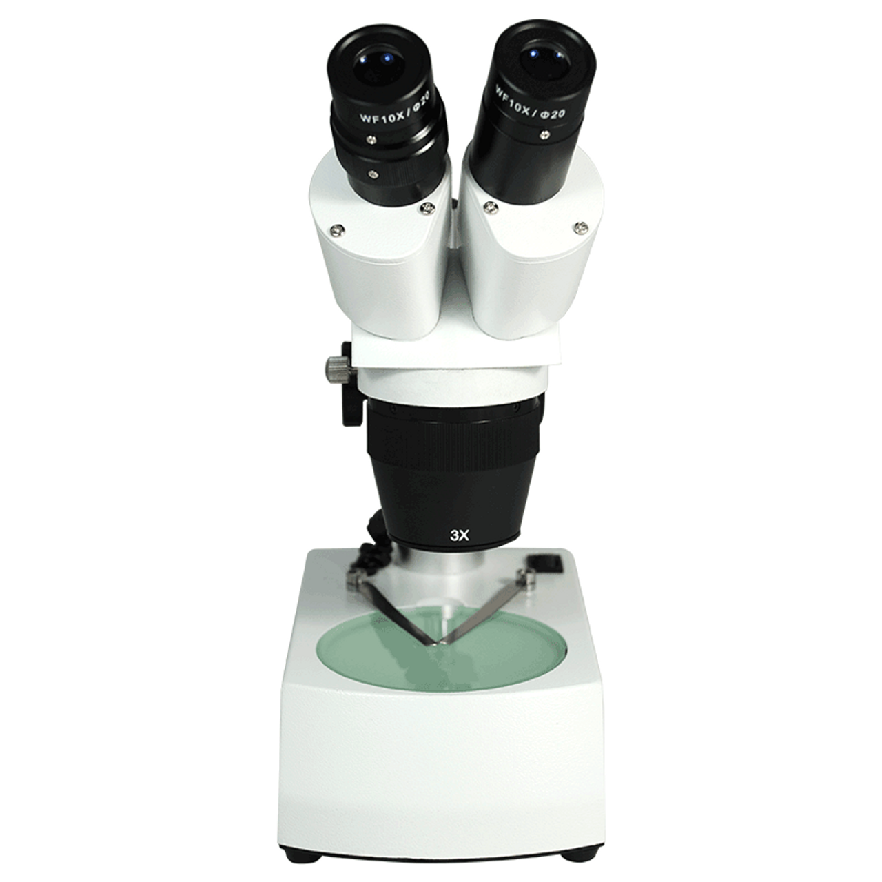 Microscopy Techniques for Observing White Blood Cells
To examine white blood cells under a microscope, specific techniques and tools are crucial. There are several microscopy methods each having its merits.
Brightfield Microscopy is the standard and most accessible technique. It shines light through the sample and works best with stained cells, enhancing the visibility of the white blood cells.
Phase-contrast Microscopy is helpful for viewing cells without staining. This technique amplifies the contrast in transparent samples, like unstained living cells. It’s ideal for watching white blood cells’ behavior in real-time.
Fluorescence Microscopy uses high-intensity light to illuminate dyed components of the cells. It’s highly specific and can locate particular elements in the cells, such as proteins or other markers.
Differential Interference Contrast (DIC) Microscopy gives a 3D-like image. It utilizes polarized light to create contrast and is great for live, unstained cells. This method highlights the detailed structures of white blood cells.
Confocal Microscopy scans the cells with a laser. It captures sharp images, slicing through the sample. This technique is excellent for detailed study of cell parts.
For accurate observations, clean slides, proper sample spreading, and precise staining are necessary. These steps ensure clear visibility of white blood cells under a microscope. Experts can then analyze size, shape, and other characteristics with greater accuracy. Every technique chosen depends on the purpose of the examination. For example, immune system research often requires precise localizations offered by fluorescence microscopy. In contrast, routine blood tests might use brightfield microscopy for speed and simplicity. Thus, selecting the proper microscopy technique is critical for detailed and correct analysis of white blood cells.
Preparing a Blood Sample for Microscopic Examination
Before examining white blood cells under microscope, proper preparation of the blood sample is essential. This preparation is a multi-step process that ensures the cells can be adequately observed and analyzed. The steps typically include collecting the sample, spreading it on a slide, fixing it, and applying the appropriate stains.
Collection: The first step involves drawing blood from the patient using a sterile technique. This is crucial to prevent contamination and ensure accurate results.
Spreading: Next, a small drop of blood is placed on a clean microscope slide. A spreader slide is then used to thinly distribute the blood across the surface, creating a ‘blood smear’.
Fixing: Once the smear is made, the sample is often ‘fixed’ to the slide. This process preserves the blood cells’ structure and prevents them from degrading or changing shape.
Staining: Finally, specific stains are applied to the slide to distinguish the cells. Stains like Wright’s or Giemsa are commonly used because they highlight the white blood cells’ characteristics. Different stains might be chosen based on the type of microscope technique to be used.
The quality of the blood sample preparation is critical in analyzing white blood cells under microscope. Well-prepared slides allow for the proper identification of different WBC types and the detection of any abnormal cells or irregularities. These steps form the basis of much of the subsequent analysis and diagnosis. Each step must therefore be performed with care and precision, ensuring that when the sample is finally placed under the microscope, it yields clear and accurate insights into the patient’s immune health.
 Identifying and Differentiating White Blood Cells Under the Microscope
Once a blood sample is prepared, the critical task is to identify and differentiate the various types of white blood cells under the microscope. Accurate identification is vital for diagnosis and treatment planning. Here’s how technicians and health professionals perform this intricate task:
Size and Shape: One of the first aspects examined is the size and shape of the cells. Different WBCs have distinctive shapes that can be recognized under microscopic analysis.
Nucleus Features: The shape and size of the nucleus is a telling feature. Neutrophils have a multi-lobed nucleus, while lymphocytes possess a large, round nucleus.
Granule Presence: Some white blood cells contain granules. Eosinophils are notable for their red-orange granules, which are easily spotted.
Staining Patterns: The type of stain used can also help differentiate between WBC types. For example, basophils stain dark blue with standard hematological stains.
Cell Count: The proportion of each white blood cell type can reveal information about current immune responses. A higher than normal count of a particular WBC type could indicate specific health issues.
Experts use these characteristics to identify and count different white blood cells under microscope. These counts provide a ‘differential’, which is an essential part of a complete blood count test. By comparing the differential with normal values, doctors can diagnose infections, allergies, or diseases like leukemia.
Identifying WBCs and noting their characteristics takes skill and careful attention to detail. Technologists often undergo extensive training to perform these tests accurately. With practice, they become proficient at recognizing subtle differences that could have significant implications for a patient’s health.
Common Findings and Abnormalities in White Blood Cells
When examining white blood cells under microscope, certain patterns and irregularities often emerge. These can be signs of normal immune responses or indications of health issues. Knowing what to look for is key for diagnosis and treatment. Here are some common findings and abnormalities seen in these cells:
Normal Range Variances: Typically, each white blood cell type falls within a certain range in healthy individuals. Small deviations from these ranges can be normal, depending on the person’s age, gender, and overall health.
Infections and Inflammation: A high white blood cell count usually suggests an active infection or inflammation. The body produces more cells to fight the invader.
Autoimmune Diseases: In conditions like lupus, the immune system attacks the body itself. This can lead to variations in white blood cell counts and appearances.
Allergic Reactions: Eosinophils increase in number during allergic reactions. They release substances that combat allergens but also cause inflammation and discomfort.
Blood Disorders: Diseases like leukemia cause abnormal white blood cell production, leading to unusual counts and immature cells in the bloodstream.
Stress Response: Physical or emotional stress can temporarily boost white blood cell levels. This is part of the body’s natural response to stressors.
These findings highlight the importance of observing white blood cells under microscope. They assist experts in determining an individual’s immune status and diagnosing potential disorders. The precise identification of abnormalities is essential for effective medical intervention.
 Advances in Microscopy and Future Directions in Hematology Research
Advancements in microscopy have transformed how we observe white blood cells under a microscope. These breakthroughs in imaging offer deeper insights into hematology, the branch of medicine related to blood. They enable us to reveal minute details of white blood cells that were not previously visible. Their impact is vast, affecting diagnosis, research, and the understanding of immune reactions.
Digital Microscopy is one of the latest innovations. It allows storing and analyzing images on a computer. This makes it easier for researchers to share findings and collaborate globally. It can also improve the accuracy of cell counts and differentiations through software analysis.
Live-cell Imaging techniques have evolved, allowing for real-time observation of white blood cells’ activities. Watching these cells interact with pathogens helps us understand immune responses better.
High-throughput Microscopy is a method that automates the analysis of large numbers of cells. It speeds up testing and provides a wealth of data, useful for extensive research studies.
Artificial Intelligence and Machine Learning are set to revolutionize microscopy. With AI, we could see systems that automatically identify and classify different types of blood cells. They might even predict changes in the immune system before they become apparent.
Looking ahead, researchers are excited about the possibilities. Nanotechnology could lead to even higher resolution imaging. We might develop tools to visualize the interactions at a molecular level. This could uncover new aspects of immune system function and disease mechanics. With these advances, the future of white blood cells under microscope study is bright. It holds promise for significant developments in the diagnosis and treatment of blood-related diseases.
The Future of White Blood Cells Under Microscope Analysis
Integration of Advanced Technologies
The future of white blood cell analysis under microscope lies in the integration of advanced technologies like AI, machine learning, and high-resolution digital imaging. These advancements will further enhance the accuracy, speed, and efficiency of diagnosing and monitoring health conditions.
Personalized Medicine
As personalized medicine continues to evolve, the detailed analysis of white blood cells under microscope will play a significant role in tailoring treatments to individual patients. Understanding the specific types and quantities of white blood cells can guide targeted therapies and improve patient outcomes.
Enhanced Research Capabilities
Future research will benefit from more sophisticated microscopy techniques, enabling deeper insights into the functions and interactions of white blood cells. This enhanced understanding will contribute to breakthroughs in immunology and the development of innovative medical treatments.
 Conclusion
In conclusion, white blood cells under microscope provide invaluable insights into our immune health and overall well-being. By understanding the different types of white blood cells, their functions, and how to observe them accurately under a microscope, we can better diagnose and monitor various health conditions. Advances in microscopy technology continue to enhance our ability to study these vital cells, paving the way for improved diagnostics, personalized treatments, and groundbreaking research in immunology.
Embracing the detailed observation of white blood cells under microscope not only contributes to individual health management but also drives advancements in medical science. Whether you are a healthcare professional, a researcher, or someone interested in the complexities of the human body, the study of white blood cells under microscope offers a fascinating glimpse into the mechanisms that protect and sustain our lives. Stay informed, stay curious, and appreciate the microscopic warriors that defend our health every day.