The microscopic realm is brimming with extraordinary life forms, and one of the most intriguing among them is the amoeba. Observing an amoeba under microscope offers a captivating glimpse into the complexity and beauty of single-celled organisms. Whether you’re a student, a biology enthusiast, or just curious about the microscopic world, understanding amoebas through microscopy can enhance your appreciation of life’s diversity. In this comprehensive article, we delve into the characteristics, behavior, and significance of amoebas, all while guiding you on how to effectively observe them under a microscope.
 What is an Amoeba?
An amoeba is a single-celled organism often found in water and soil. These microscopic beings are part of the Protista kingdom. They have a flexible shape and move using pseudopods. Pseudopods are temporary arms that an amoeba projects forward to crawl along surfaces. The body of an amoeba is very simple. It lacks a fixed form and changes shape as it moves. Amoebas play a key role in their ecosystems. They consume bacteria and other small particles through a process called phagocytosis. This is when the amoeba surrounds and engulfs its prey with its body. Amoebas reproduce by splitting into two. This process is known as binary fission. These organisms are fascinating to study under a microscope. They can offer insights into the building blocks of life and the evolution of complex organisms. Looking at an amoeba under a microscope reveals the basic life activities of all cells.
The Structure of Amoeba
The structure of an amoeba is simple yet intricate. Unlike multi-cellular organisms, the amoeba’s body consists of a single cell performing all life functions. Despite its simplicity, understanding the structure of an amoeba under a microscope gives us incredible insight into cellular biology.
Cellular Components and Functions
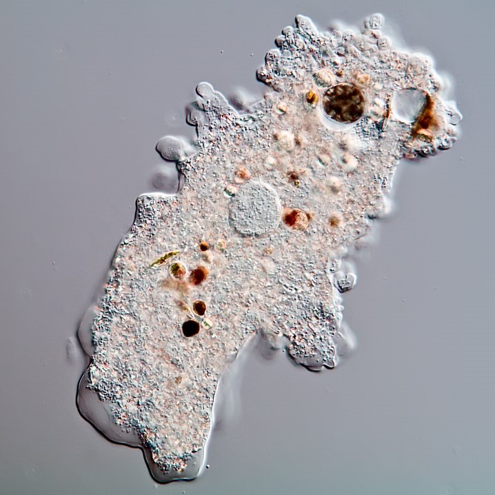
When viewed through a microscope, the amoeba reveals various cellular components, each with a specific function. The most visible feature is the cell membrane, a flexible boundary that holds the cell contents and controls entry and exit of substances. Inside the membrane lies the cytoplasm, a jelly-like substance where most cellular activities occur. Suspended in the cytoplasm, the nucleus can be identified, which houses DNA and regulates growth and reproduction.
The amoeba also contains vacuoles, which are small sacs used for storage and digestion. Food vacuoles store ingested food particles, while contractile vacuoles expel excess water to maintain osmotic balance. The amoeba’s iconic pseudopods are extensions of the cell membrane and cytoplasm. They allow the amoeba to move and feed by enveloping its prey.
These basic cellular components work in harmony, ensuring the amoeba’s survival, growth, and reproduction. Each function serves as a fundamental example of cellular life, making the amoeba a perfect specimen to observe under a microscope. With amoeba under a microscope, scientists and students alike can witness the intricacies of life’s smallest units.
 Observing Amoeba Under a Microscope
To understand the life and structure of an amoeba, observing these organisms under a microscope is crucial. The process brings the invisible world of single-celled organisms to life. It grants us a microscopic view of the complex activities carried out by these simple beings.
Preparing the Specimen
Before observing an amoeba under a microscope, proper specimen preparation is essential. Collect a sample from a water source. Place a drop on a glass slide. Use a cover slip to avoid air bubbles. The amoeba must be alive to observe its movements and feeding habits effectively. Therefore, samples should be fresh and examined promptly.
In the lab, staining can be employed to make cell structures more visible. However, it’s important to note that stains may kill the amoeba. Therefore, unstained wet mounts are preferable for observing living amoebas. Adjust the microscope to a lower magnification to locate the amoeba. Then, switch to a higher magnification for a detailed view.
Identifying Amoeba Among Other Microorganisms
Finding an amoeba under a microscope can be tricky due to their transparent bodies and slow movement. Look for irregular shapes that change over time. This is indicative of an amoeba’s pseudopod activity. Distinct from rigid microorganisms, an amoeba’s flexibility is a telltale sign.
Scan for active feeding behavior. Amoebas engulf particles, creating visible food vacuoles. These vacuoles are a clear indicator of amoeba presence. Additionally, amoebas often appear larger than many other single-celled organisms in the sample. By familiarity with these basic identification tips, an amoeba under a microscope can stand out amidst the multitude of microorganisms present in the sample.
The Life Cycle of Amoebas
Understanding the life cycle of amoebas adds depth to our study of these microorganisms. The amoeba life cycle is a captivating process, simple yet efficient, ensuring the continuation of the species.
Reproduction Process
Amoebas reproduce asexually through binary fission, a straightforward and fascinating process to observe under a microscope. In binary fission, the amoeba’s nucleus divides, replicating its genetic material. Following nuclear division, the cytoplasm separates, creating two identical amoebas from the original one. This method of reproduction allows for rapid population growth in suitable environments.
Amoeboid reproduction does not involve mating or exchange of genetic material. This means each offspring is genetically identical to the parent, a clone. Binary fission may occur when the amoeba has ingested sufficient food and is in an ideal state of health. The process is not seasonally bound and can happen multiple times, contributing to their persistence in their natural habitats.
Observing this process under a microscope reveals the incredible autonomy of single-celled organisms like amoebas. They embody the essence of life, thriving independently, without the complexity of multi-organ systems. Their reproductive efficiency plays a critical role in their survival and proliferation.
 Amoeba in their Natural Habitat
Amoebas are more than microscopic wonders; they are vital to their ecosystems.
Role in the Ecosystem
In their natural habitats, amoebas serve important roles. Generally found in water or moist soil, they help to break down organic material. This recycling of nutrients turns waste back into resources for other organisms. Additionally, amoebas control bacterial populations by consuming them. This process checks bacterial overgrowth, which could otherwise harm the ecosystem’s balance.
By feeding on bacteria, amoebas indirectly support larger organisms. The consumed bacteria could have otherwise competed with these larger beings for nutrients. Also, amoebas can be food sources for other microorganisms and small aquatic animals. Thus, they are a key link in the food chain. Furthermore, their presence indicates a healthy environment, as they thrive in unpolluted water.
Studying amoebas provides insights into water quality and the health of ecosystems. Identifying the diversity and number of amoebas can help gauge the level of pollution. To sum up, amoebas are not only intriguing subjects under a microscope but also essential to ecological health and balance.
The Importance of Amoebas in Research
Amoebas serve as critical tools in the field of biological and medical research. Their relatively simple structure and life processes provide a groundwork for understanding more complex organisms. When scientists examine an amoeba under a microscope, they gain valuable insights into cellular behavior, genetics, and life cycle dynamics.
The diverse functions & mechanisms present within these single-celled creatures reflect foundational aspects of life. For example, the process of phagocytosis in amoebas is akin to the mechanisms by which human cells combat pathogens. By studying these processes in amoebas, researchers can draw parallels to human biology, potentially leading to breakthroughs in treatments and medicine.
Furthermore, impressive as it seems, amoebas are a model organism for studying genetic regulation and expression. Without the complexities of multi-cellular organisms, amoebas offer a clearer view of how genetic material directs all aspects of life – growth, reproduction, and adaptation.
In toxicology, amoebas are used to assess environmental pollutants. Their reaction to toxic substances provides quick and reliable data on the pollutants’ potential impact on life forms. They are sensitive to changes in their surroundings, making them great bio-indicators.
Lastly, amoebas’ asexual reproductive behavior – binary fission – helps in research about population dynamics and evolutionary biology. By observing amoebas, scientists study how populations grow and survive without the genetic variety presented by sexual reproduction.
In summary, amoebas, while tiny, are mighty in research. They are windows into the cell’s world, offering a vast wealth of knowledge to those who study them under a microscope.
 Amoeba Movement and Behavior
To grasp how amoebas interact with their world, it’s vital to delve into their movement and behavior. These fascinating microorganisms display unique locomotion abilities, which have been subjects of study for many years.
Understanding Amoeboid Movement
Amoeboid movement is the hallmark of an amoeba, giving these organisms their name. This type of motion involves the expansion and retraction of pseudopods. Here’s a step-by-step guide to how it works:
- The amoeba first extends a portion of its cell body, creating a pseudopod.
- The cytoplasm flows into this extension, thrusting the rest of the cell forward.
- The amoeba’s cell membrane anchors to a surface, allowing the cell to pull itself onward.
- As it moves, the amoeba may change directions by forming new pseudopods, retracting old ones.
- Through this method, the amoeba can navigate its environment, searching for food and escaping predators.
Watching an amoeba under a microscope is like witnessing a ballet of cellular dynamics. Each movement is a response to the amoeba’s immediate surroundings. They adjust their course to optimize feeding, avoid harsh conditions, and react to stimuli. Their behavior under a microscope provides a basic understanding of how cells interact with their environment, showcasing life at its most fundamental level. To ensure observations are accurate, amoeba under microscope samples must be fresh and lively. This enables clear visibility of their behavior and interaction with other microorganisms.
Encountering Amoebas: Safety and Health Concerns
While studying amoebas under a microscope is safe, encountering them in natural habitats can pose risks. Most amoebas are harmless, but some species can affect health adversely. For example, the Naegleria fowleri, often known as the “brain-eating amoeba,” can cause severe infections if it enters the human body through the nasal passages. This can lead to a rare but deadly condition known as primary amoebic meningoencephalitis (PAM).
Here are some safety measures to consider:
- Avoid swimming or diving in warm, stagnant waters where amoebas may thrive.
- Use nose clips or keep your head above water in hot springs or untreated pools.
- When cleaning fish tanks or handling soil, wear protective gloves to avoid direct contact.
- Always boil or filter water obtained from lakes or rivers before drinking.
Education on amoebas under a microscope should include their potential health risks. Quick medical attention can be life-saving if one suspects exposure to harmful amoebas. Always observe safety practices in environments where amoebas are present. By doing so, we can appreciate the remarkable nature of amoebas without compromising our health.
 Future Directions in Amoeba Research
Advanced Imaging Techniques
The future of amoeba research lies in the development of more advanced imaging techniques. Technologies such as super-resolution microscopy and live-cell imaging will enable scientists to observe amoebas with unprecedented detail and fidelity. These advancements will uncover new aspects of amoebic biology and behavior, pushing the boundaries of our understanding.
Synthetic Biology
Exploring synthetic biology with amoebas opens new avenues for research and applications. By engineering amoebas with specific genetic traits, researchers can create customized organisms for purposes such as drug delivery, environmental remediation, and bioengineering. This innovative approach holds great promise for future scientific and technological breakthroughs.
Collaborative Research Initiatives
Collaborative research initiatives that bring together experts from various fields can accelerate discoveries about amoebas. Interdisciplinary collaborations between biologists, geneticists, environmental scientists, and engineers will foster a holistic approach to studying amoebas, leading to more comprehensive insights and impactful applications.
Conclusion: The Endless Fascination of Amoeba Under Microscope
In conclusion, observing an amoeba under microscope unveils a world of complexity and beauty that exists beyond our naked eye. From understanding their cellular structures and behaviors to appreciating their role in ecosystems and human health, amoebas offer endless fascination and valuable scientific knowledge. By mastering the techniques for preparing and observing amoebas, and by staying informed about the latest research and advancements, enthusiasts and researchers alike can continue to explore and appreciate these remarkable single-celled organisms. Embrace the opportunity to delve into the microscopic universe, and let the study of amoebas inspire a deeper connection with the intricate and diverse forms of life that share our world.