Understanding the microscopic structure of hyaline cartilage under microscope reveals essential details about its role in the human body. This intricate tissue plays a crucial part in joint function, growth, and repair. By examining hyaline cartilage under a microscope, scientists and medical professionals gain valuable insights into its composition, health, and response to various conditions. In this comprehensive article, we will delve into the fascinating world of hyaline cartilage, exploring its microscopic features, functions, and the significance of studying it at such a detailed level.
 What is Hyaline Cartilage?
Hyaline cartilage is the most common type of cartilage found in the body. It provides flexibility and support to various structures, including joints, rib cage, nose, and airways. This type of cartilage is characterized by its glossy, bluish-white appearance and smooth texture, making it essential for facilitating smooth movements and reducing friction in joints.
Structure of Hyaline Cartilage
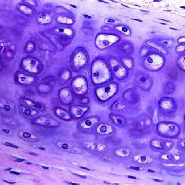
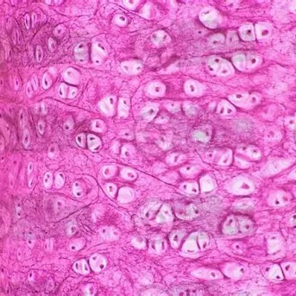
When examining hyaline cartilage under microscope, several key structural components become evident. The tissue is composed of chondrocytes, which are specialized cells responsible for producing and maintaining the cartilage matrix. These cells reside in small spaces called lacunae within the extracellular matrix, which consists of collagen fibers, proteoglycans, and water.
Extracellular Matrix Composition
The extracellular matrix of hyaline cartilage plays a vital role in its function and resilience. Collagen type II is the primary protein fiber, providing tensile strength and maintaining the cartilage’s shape. Proteoglycans, mainly aggrecan, attract and retain water molecules, contributing to the cartilage’s ability to withstand compressive forces. This combination of collagen and proteoglycans creates a resilient and flexible structure that supports various bodily functions.
Functions of Hyaline Cartilage
Hyaline cartilage serves multiple purposes in the body, each crucial for maintaining overall health and mobility. Let’s explore the primary functions of this remarkable tissue.
Joint Support and Movement
In joints, hyaline cartilage forms the articular cartilage that covers the ends of bones. This smooth surface allows for frictionless movement, enabling joints to move smoothly and efficiently. Additionally, hyaline cartilage acts as a shock absorber, distributing mechanical stress and preventing bone damage during activities like walking, running, and jumping.
Respiratory System Function
Hyaline cartilage is also present in the respiratory system, where it forms structures like the trachea and bronchi. In these airways, hyaline cartilage maintains the shape and openness of the tubes, ensuring unobstructed airflow. This flexibility is crucial for breathing, as it allows the airways to expand and contract with each breath.
Growth and Development
During fetal development and childhood, hyaline cartilage serves as a precursor to bone formation in a process known as endochondral ossification. The growth plates, or epiphyseal plates, are composed of hyaline cartilage, which gradually transforms into bone as children reach maturity. This transition is essential for the proper growth and lengthening of long bones.
Examining Hyaline Cartilage Under Microscope
Analyzing hyaline cartilage under microscope provides a deeper understanding of its intricate structure and function. Microscopic examination reveals details that are not visible to the naked eye, allowing for precise assessments of cartilage health and pathology.
Histological Features
When hyaline cartilage is viewed under a microscope, several distinct histological features become apparent. The dense network of collagen fibers forms a matrix that supports the chondrocytes. These cells are evenly distributed within the matrix, maintaining the tissue’s integrity and facilitating nutrient exchange. The smooth, glassy appearance of the cartilage is due to the highly organized arrangement of its cellular and extracellular components.
Types of Microscopic Staining
Different staining techniques enhance the visibility of various components of hyaline cartilage under microscope. Hematoxylin and eosin (H&E) staining highlights the cellular structures, making chondrocytes and lacunae easily identifiable. Safranin O staining is used to detect proteoglycans, as it imparts a red color to these molecules, allowing for the assessment of matrix composition and health.
Microscopic Pathologies
Studying hyaline cartilage under a microscope can reveal pathological changes that indicate diseases or injuries. For instance, in osteoarthritis, microscopic examination shows the breakdown of cartilage matrix, loss of chondrocytes, and increased inflammation. These changes compromise the cartilage’s ability to function properly, leading to joint pain and decreased mobility.
 Importance of Microscopic Analysis
Microscopic analysis of hyaline cartilage is essential for diagnosing and understanding various medical conditions. By examining the tissue at a cellular level, healthcare professionals can identify early signs of degeneration, inflammation, and other abnormalities that may require intervention.
Early Diagnosis of Joint Diseases
Detecting joint diseases like osteoarthritis and rheumatoid arthritis at an early stage can significantly improve treatment outcomes. Microscopic examination allows for the identification of subtle changes in cartilage structure and composition before severe symptoms develop, enabling timely medical intervention and management.
Research and Development
Microscopic studies of hyaline cartilage contribute to the advancement of biomedical research. Understanding the cellular and molecular mechanisms underlying cartilage health and disease paves the way for developing innovative therapies and regenerative medicine approaches. This knowledge is crucial for designing treatments that can restore or enhance cartilage function.
Tissue Engineering and Regenerative Medicine
The insights gained from examining hyaline cartilage under microscope are instrumental in the field of tissue engineering. Scientists use this information to develop scaffolds and biomaterials that mimic the natural cartilage environment, promoting the growth and differentiation of chondrocytes. These advancements hold promise for repairing damaged cartilage and improving the quality of life for individuals with joint disorders.
Techniques for Microscopic Examination
Several techniques are employed to study hyaline cartilage under microscope, each offering unique advantages for detailed analysis.
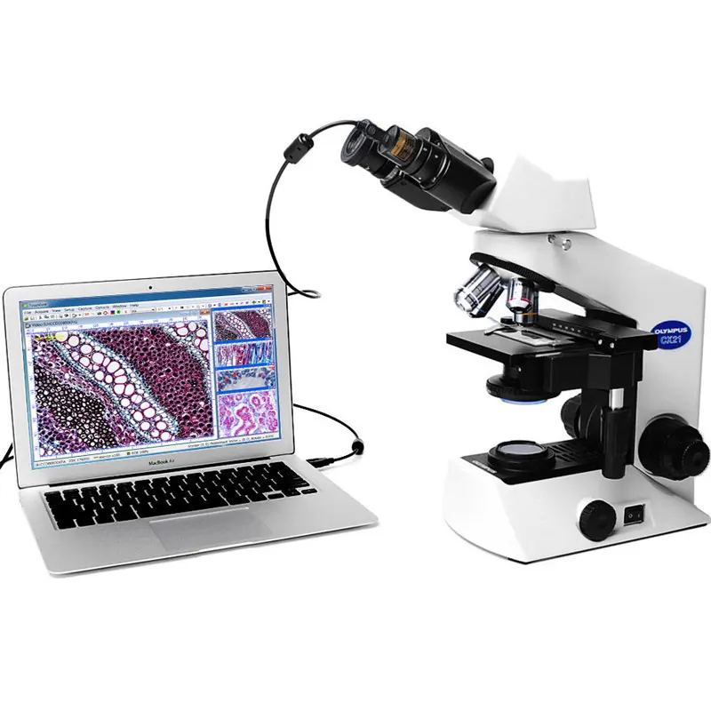
Light Microscopy
Light microscopy is the most common method used to examine hyaline cartilage. By using various staining techniques, light microscopes can reveal the cellular and extracellular structures of the tissue. This method is widely accessible and provides sufficient resolution for most clinical and research purposes.
Electron Microscopy
For even more detailed visualization, electron microscopy can be used to study hyaline cartilage at a nanoscopic level. Transmission electron microscopy (TEM) and scanning electron microscopy (SEM) offer high-resolution images of the cartilage’s ultrastructure, including the arrangement of collagen fibers and the morphology of chondrocytes. These techniques are invaluable for advanced research applications.
Confocal Microscopy
Confocal microscopy allows for the three-dimensional visualization of hyaline cartilage. By using fluorescent markers, this technique can highlight specific components within the tissue, providing insights into cellular interactions and matrix organization. Confocal microscopy is particularly useful for studying live cartilage samples and dynamic processes.
 Factors Affecting Hyaline Cartilage Health
Several factors influence the health and integrity of hyaline cartilage. Understanding these factors is crucial for maintaining cartilage function and preventing degenerative diseases.
Mechanical Stress and Trauma
Excessive mechanical stress or trauma can damage hyaline cartilage, leading to wear and tear. Activities that involve repetitive motion or high-impact forces, such as running or jumping, can accelerate cartilage degeneration. Proper training, protective gear, and ergonomic practices can help mitigate these risks.
Aging
As individuals age, hyaline cartilage naturally undergoes degeneration. The loss of chondrocytes, decreased synthesis of extracellular matrix components, and increased inflammation contribute to the decline in cartilage quality. Age-related changes increase the susceptibility to joint diseases like osteoarthritis.
Inflammation and Immune Response
Chronic inflammation and immune responses can adversely affect hyaline cartilage. Inflammatory cytokines and enzymes released during autoimmune conditions or infections can degrade the cartilage matrix and inhibit repair processes. Managing inflammation through medication and lifestyle changes is essential for preserving cartilage health.
Nutritional Deficiencies
Adequate nutrition is vital for maintaining healthy hyaline cartilage. Essential nutrients like vitamin C, vitamin D, calcium, and omega-3 fatty acids support cartilage repair and regeneration. A balanced diet rich in these nutrients helps sustain the structural integrity and function of hyaline cartilage.
Genetic Factors
Genetics play a significant role in determining the susceptibility to cartilage-related conditions. Certain genetic mutations can affect the synthesis and organization of cartilage matrix components, increasing the risk of degeneration and disease. Understanding genetic predispositions can aid in early detection and personalized treatment strategies.
Therapeutic Approaches for Cartilage Damage
Various therapeutic approaches aim to repair or regenerate damaged hyaline cartilage, improving joint function and reducing pain.
Conservative Treatments
Conservative treatments focus on managing symptoms and slowing the progression of cartilage damage. These include:
- Physical Therapy: Strengthening the muscles around the joints can reduce stress on hyaline cartilage.
- Medications: Nonsteroidal anti-inflammatory drugs (NSAIDs) alleviate pain and inflammation.
- Lifestyle Modifications: Weight management and low-impact exercises help preserve cartilage health.
Surgical Interventions
In cases of significant cartilage damage, surgical interventions may be necessary. Common procedures include:
- Microfracture Surgery: Stimulates the growth of new cartilage by creating small fractures in the bone.
- Autologous Chondrocyte Implantation (ACI): Transplants healthy chondrocytes to the damaged area to promote regeneration.
- Osteochondral Autograft Transfer System (OATS): Transfers healthy cartilage from a non-weight-bearing area to the damaged site.
Regenerative Medicine
Regenerative medicine offers promising solutions for hyaline cartilage repair. Techniques such as stem cell therapy and tissue engineering aim to restore the natural structure and function of cartilage. These advanced approaches hold potential for long-term regeneration and improved outcomes for individuals with cartilage damage.
 Future Directions in Cartilage Research
Ongoing research continues to uncover new insights into hyaline cartilage, driving advancements in diagnosis, treatment, and prevention of cartilage-related conditions.
Biomaterials and Scaffold Development
Developing innovative biomaterials and scaffolds can enhance cartilage regeneration efforts. These materials provide a supportive environment for chondrocytes and promote the formation of new extracellular matrix
Advances in Imaging Technologies
Improved imaging technologies offer more precise and detailed visualization of hyaline cartilage. Innovations in MRI and ultrasound techniques enhance the ability to detect early cartilage changes and monitor treatment efficacy, leading to better clinical outcomes.
Gene Therapy and Molecular Approaches
Gene therapy and molecular approaches aim to address the root causes of cartilage degeneration. By targeting specific genes and molecular pathways, these techniques can promote cartilage regeneration and prevent further damage. Research in this area holds promise for developing targeted and effective treatments.
Personalized Medicine
Personalized medicine tailors treatments based on an individual’s genetic makeup and specific cartilage conditions. This approach ensures that therapies are more effective and minimize adverse effects, improving overall patient outcomes. Personalized strategies are becoming increasingly important in the management of cartilage-related diseases.
Real-World Applications and Case Studies
Examining hyaline cartilage under microscope has numerous real-world applications, enhancing our understanding and treatment of various conditions.
Osteoarthritis Diagnosis and Management
Microscopic analysis of hyaline cartilage has revolutionized the diagnosis and management of osteoarthritis. By identifying early cartilage changes, healthcare providers can implement preventive measures and tailored treatments to slow disease progression and alleviate symptoms.
Sports Medicine and Injury Prevention
In sports medicine, understanding the microscopic structure of hyaline cartilage helps in designing better training programs and injury prevention strategies. Athletes benefit from targeted interventions that protect cartilage health, enhancing performance and reducing the risk of joint injuries.
Veterinary Medicine
Hyaline cartilage studies are not limited to humans; they also play a crucial role in veterinary medicine. Understanding cartilage structure and function in animals helps in diagnosing and treating joint disorders in pets and livestock, improving their quality of life and productivity.
Biomedical Engineering
Biomedical engineers utilize insights from microscopic analysis of hyaline cartilage to develop artificial cartilage and joint replacement materials. These advancements provide viable alternatives for individuals with severe cartilage damage, restoring joint function and mobility.
Challenges in Studying Hyaline Cartilage Under Microscope
Despite significant advancements, studying hyaline cartilage under microscope presents several challenges that researchers continue to address.
Limited Regenerative Capacity
Hyaline cartilage has a limited ability to regenerate, making it difficult to study and repair. Understanding the factors that influence cartilage regeneration is essential for developing effective therapies.
Complex Tissue Architecture
The intricate structure of hyaline cartilage poses challenges for microscopic analysis. Ensuring accurate and detailed visualization requires advanced techniques and expertise, which can be resource-intensive.
Variability in Cartilage Composition
Cartilage composition can vary based on location, age, and health status. This variability complicates the study of hyaline cartilage, as it requires careful consideration of different factors that influence its structure and function.
Accessibility of Samples
Obtaining high-quality cartilage samples for microscopic analysis can be challenging. Invasive procedures are often required to collect tissue samples, which can limit the availability of specimens for research.
 Practical Tips for Researchers
For those conducting research on hyaline cartilage under microscope, here are some practical tips to enhance accuracy and efficiency.
Sample Preparation
Proper sample preparation is crucial for obtaining clear and detailed microscopic images. Use appropriate fixation, embedding, and sectioning techniques to preserve the tissue’s structure and composition.
Choosing the Right Staining Method
Select staining methods that highlight the specific components you wish to study. For example, use H&E staining for general histological features and Safranin O for proteoglycan detection.
Utilizing Advanced Imaging Techniques
Incorporate advanced imaging techniques such as confocal microscopy and electron microscopy to gain deeper insights into cartilage structure and function. These methods provide higher resolution and more detailed views of the tissue.
Collaborative Research
Collaborate with experts in related fields, such as molecular biology and biomedical engineering, to enhance the scope and impact of your research. Interdisciplinary approaches can lead to more comprehensive and innovative findings.
Continuous Learning
Stay updated with the latest advancements in cartilage research and microscopy techniques. Participating in workshops, conferences, and training programs can help you stay ahead in the field.
Conclusion
Examining hyaline cartilage under microscope offers invaluable insights into its structure, function, and role in maintaining joint health and overall mobility. This microscopic perspective is essential for diagnosing cartilage-related conditions, advancing medical research, and developing effective treatments. By understanding the intricate details of hyaline cartilage, we can better appreciate its importance in the human body and work towards preserving its health through informed medical interventions and innovative therapies. Embracing the knowledge gained from microscopic studies ensures that we continue to improve the quality of life for individuals affected by cartilage disorders and promote advancements in biomedical science.