A kidney stone microscope plays a crucial role in modern medical diagnostics, offering detailed insights into the composition and structure of kidney stones. Understanding the functionalities and applications of a kidney stone microscope can significantly improve patient outcomes and treatment strategies. This article explores the various aspects of kidney stone microscopes, including their types, how they work, their benefits in the medical field, and tips for selecting the right microscope for clinical use. By delving into these details, healthcare professionals and medical facilities can make informed decisions to enhance their diagnostic capabilities.
 The Basics of Kidney Stones
Understanding kidney stones begins with a basic grasp of what they are. When minerals and salts in urine clump together, they may form kidney stones. These stones can range in size and shape. They may go unnoticed until they cause problems, such as pain or infection.
Definition and Types of Kidney Stones
Kidney stones are hard deposits that form in the kidneys. These deposits can develop for various reasons and consist of different combinations of minerals. The four main types are:
- Calcium stones, mostly in the form of calcium oxalate. Oxalate is a substance found in foods and is produced daily by your liver.
- Uric acid stones, which can form in people who do not drink enough fluids or who lose too much fluid, those who eat a high-protein diet, and those with gout.
- Struvite stones, typically developing in response to an infection, like a urinary tract infection.
- Cystine stones, which occur in people with a hereditary disorder that causes the kidneys to excrete too much of certain amino acids.
Causes and Risk Factors
Several factors can increase the risk of developing kidney stones. Not drinking enough water is a leading cause, as it can lead to more concentrated urine, where minerals can crystallize and stick together. Dietary factors, like high intake of salt or sugar, may also increase the chance of stone formation. Obesity, certain supplements and medications, and medical conditions such as Crohn’s disease or hyperparathyroidism can play a role as well.
Other risk factors include:
- A family or personal history of kidney stones.
- Frequent urinary infections.
- Conditions that affect how your body absorbs and uses calcium and other minerals.
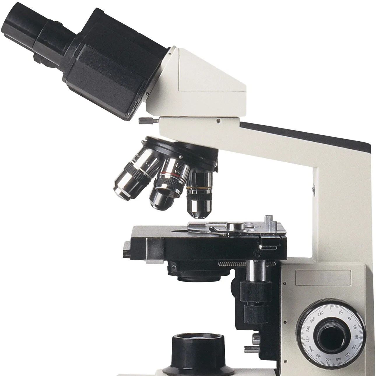
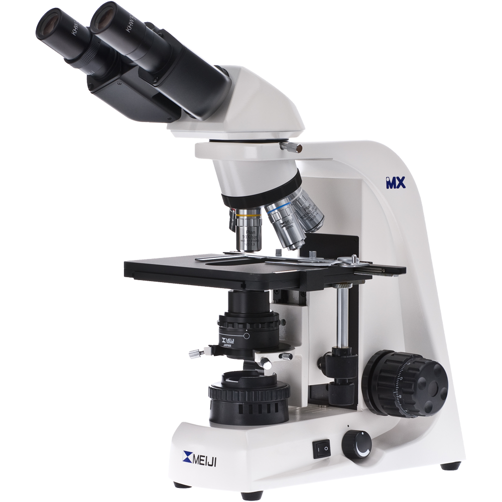
Analyzing kidney stones through a microscope, commonly referred to as ‘kidney stone microscope,’ can help in identifying their composition and type, thus guiding treatment decisions.
 Microscopic Characteristics of Kidney Stones
When viewed under a kidney stone microscope, these deposits reveal a world of complex structures.
Composition Analysis
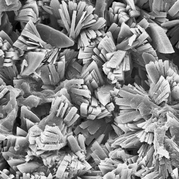
Microscopic analysis is key in understanding kidney stone composition. This involves inspecting crushed stone fragments or their dust under a microscope. Pathologists can often pinpoint the minerals present. For example, calcium stones may show clear signs of calcium oxalate. Uric acid stones, under the microscope, lack the crystalline structure seen in calcium stones.
By identifying the exact minerals, doctors can determine their potential causes. For instance, high levels of oxalate may suggest issues with a patient’s diet. Or, it could point to the liver producing too much oxalate. The microscopic exam helps tailor treatment to prevent future stones.
Crystal Structure and Types
The kidney stone microscope exposes varied crystal structures. The shape and size of these crystals can tell a lot about a stone’s makeup. Calcium oxalate stones, for example, often appear as envelope-shaped crystals. This is unlike struvite stones, which display coffin-lid shaped crystals. These structural differences also assist physicians in forming a treatment plan.
By closely identifying the type of crystals, health care providers can advise on specific dietary changes. They may also consider a particular type of medication that targets the stone’s composition. This approach not only treats the current stones but also aims to reduce the possibility of new stone formation.
Kidney Stone Diagnosis
Diagnosis of kidney stones often combines several techniques. These aim to understand size, location, and composition.
Imaging Techniques
Doctors use imaging methods to detect kidney stones. Common techniques include ultrasounds and CT (computed tomography) scans. Ultrasounds spot stones by bouncing sound waves off the kidneys. CT scans provide detailed images. They help see the stones’ exact positions in the urinary tract. X-rays are less common but can still find some types of stones. Together, these imaging tools present a clear picture of the physical presence of stones.
Role of Microscopy in Diagnosis
Microscopy adds another layer to diagnosis. After imaging spots the stones, microscopy examines their makeup. A kidney stone microscope looks at their tiny details. Experts crush stones to analyze under the microscope. They identify minerals and the structure of crystals. This process often finds what images cannot show — the stone’s composition. Knowing this helps doctors choose the best treatment plan. Microscopy plays a key role in understanding the makeup of stones, essential for tailored care.
 Treatment Options for Kidney Stones
When dealing with kidney stones, treatment aims to relieve symptoms and prevent future formations. Two main approaches include medical management and surgical interventions.
Medical Management
Medical management focuses on treating smaller stones that might pass on their own. Common strategies involve:
- Hydration: Drinking enough fluids helps flush out stones through urination.
- Pain Relievers: Over-the-counter medication eases the pain that comes with passing stones.
- Medical Expulsion Therapy: Prescribed drugs like tamsulosin relax the muscles in the ureter, helping stones pass.
- Dietary Changes: Modifying intake of certain foods reduces the risk of stone recurrence. A focus on a diet low in salt and animal proteins is often recommended.
The choice of medication may depend on the type of stone. For calcium stones, doctors might prescribe a thiazide diuretic or phosphate. For uric acid stones, allopurinol is often used to reduce uric acid levels.
Surgical Interventions
Larger stones that can’t pass on their own, or that cause bleeding, kidney damage, or ongoing urinary tract infections may require more aggressive treatment, such as:
- Shock Wave Lithotripsy (SWL): This non-invasive procedure uses shock waves to break stones into small pieces that can pass in the urine.
- Ureteroscopy: A small scope is inserted through the bladder to remove stones or break them down with a laser.
- Percutaneous Nephrolithotomy: When stones are too large for SWL or ureteroscopy, a surgical procedure removes them directly from the kidney.
Doctors choose surgical intervention based on the size, location, and composition of the kidney stone. As revealed by kidney stone microscope analysis, understanding the makeup of the stone guides the choice of the treatment type. Surgical treatments often follow medical management when conservative methods are insufficient or inappropriate for the patient’s condition.
Preventing Kidney Stones
Preventing kidney stones is possible with key dietary and lifestyle changes. Addressing these factors can reduce the risk of stone formation.
Dietary Adjustments
To prevent kidney stones, adjustments to your diet are crucial. Here are some tips:
- Reduce Salt: High sodium intake can cause calcium buildup, which may lead to stones.
- Limit Oxalate-Rich Foods: Foods like spinach and nuts have oxalates that bind with calcium in urine.
- Stay Hydrated: Drinking plenty of fluids, especially water, dilutes the substances that form stones.
- Moderate Protein: Lower animal protein consumption to reduce uric acid stone risks.
- Calcium Intake: Get the right amount of calcium from foods, not supplements, to inhibit stone formation.
By using a kidney stone microscope, doctors can identify if specific dietary habits contribute to stone formation. This informs tailored dietary advice.
Lifestyle Modifications
Lifestyle choices play a significant role in preventing kidney stones. Here’s what can help:
- Regular Exercise: Staying active helps prevent obesity, which is linked to stone formation.
- Hydration Habits: Make a habit of drinking water throughout the day, not just when thirsty.
- Weight Management: Achieve and maintain a healthy weight to reduce the risk of stones.
- Avoid Excessive Caffeine: Too much caffeine can increase calcium in urine, leading to stones.
Following these guidelines helps your body maintain a balance, lessening the chances of kidney stones. Your healthcare provider may use a kidney stone microscope to ensure your preventive measures align with the type of stones you’re inclined to develop.
 Recent Advances in Kidney Stone Analysis
The field of kidney stone analysis is continuously evolving. New techniques are enhancing the precision and ease with which doctors can investigate and treat kidney stones.
Technological Innovations
Technological progress has led to significant improvements in kidney stone analysis. Advanced imaging technologies, like dual-energy CT scans, can now differentiate between stone types better than traditional scans. This reduces the need for a kidney stone microscope in some cases. Innovations in endoscopic equipment have also allowed for better visualization during surgical procedures. Further, lasers with finer control are making the fragmentation of stones more efficient and less invasive.
In the laboratory, innovative devices like digital microscopes have become more sophisticated. They are providing higher resolution images for a more detailed analysis of kidney stone crystal structure. Additionally, new lab techniques, such as Fourier-transform infrared spectroscopy, are being used to identify stone composition with greater accuracy. These technological advancements lead to personalized treatment plans based on precise stone analysis.
Research Findings and Implications
Recent studies have shed light on the genetic and environmental factors contributing to kidney stone formation. Research findings have underscored the importance of genetic predispositions in stone development, pointing to new potential targets for prevention and therapy. Investigations into dietary influences have resulted in refined guidelines for nutrition, emphasizing the role of specific foods and supplements in kidney stone prevention.
These research efforts have significant implications. They enable healthcare providers to give more accurate advice on preventing recurrence, based on individual risk factors. Moreover, understanding the interplay between genetics and environment helps in creating awareness of the potential for stone development, allowing early intervention. This keeps treatment tailored and effective, improving patient outcomes.
Ongoing studies and their results are essential, as they refine existing knowledge and inform the future of kidney stone treatment.
Case Studies: Microscopic Examinations and Outcomes
The use of the kidney stone microscope has remarkable stories of success and lessons from more complex cases. These real-world examples show the value of detailed analysis in managing kidney stones effectively.
Success Stories
Many patients have benefited from microscopic examinations. Accurate analysis of kidney stones often leads to successful, non-invasive treatments. For example, pinpointing a high oxalate composition spurred dietary changes that dissolved the stones, and the patient avoided surgery. Another success involved a patient with recurrent stones. The microscope revealed a unique crystal structure, leading to a tailored medical therapy that stopped further formations. These cases demonstrate how understanding the microscopic details can lead to triumphant outcomes.
Learning from Complex Cases
However, not all cases are straightforward. Some patients present with stones that have more complex makeup. Microscopy has shown mixtures of different crystal types that complicate treatment. In these instances, doctors combine medication with dietary adjustments to tackle the issue. For tough cases that do not respond well to medical management, a careful study of the microscopic features guided doctors to choose less common, but more effective, surgical techniques. Learning from these complex cases helps refine treatment protocols and improve future care plans. Each case advances the knowledge in the field, making the kidney stone microscope an indispensable tool in the fight against kidney stones.
Advancements in Kidney Stone Microscopy
The field of kidney stone microscopy continues to evolve, incorporating technological advancements that enhance diagnostic capabilities and research opportunities.
Automation and AI Integration
Integrating automation and artificial intelligence enhances the efficiency and accuracy of kidney stone analysis:
- Automated Imaging: Streamlines the imaging process, reducing manual intervention and increasing throughput.
- AI Analysis: Utilizes machine learning algorithms to analyze stone characteristics, providing faster and more accurate diagnoses.
- Data Management: AI helps in organizing and interpreting large datasets, supporting comprehensive research and clinical studies.
Enhanced Imaging Techniques
New imaging techniques improve the depth and clarity of kidney stone analysis:
- High-Resolution Imaging: Advances in sensor technology offer sharper and more detailed images.
- 3D Imaging: Provides three-dimensional views of stone structures, aiding in comprehensive analysis.
- Multispectral Imaging: Combines different wavelengths of light to reveal various stone components and features.
Portable and Compact Designs
Portable kidney stone microscopes increase accessibility and convenience in diverse settings:
- Field Use: Enables on-site analysis in remote or resource-limited environments.
- Compact Models: Suitable for smaller laboratories or clinics with limited space.
- User-Friendly Interfaces: Simplified controls and lightweight designs make these microscopes easier to handle and operate.
Sustainable and Eco-Friendly Innovations
Sustainable practices are becoming increasingly important in medical equipment design:
- Energy Efficiency: Modern microscopes use less power without compromising performance.
- Eco-Friendly Materials: Incorporate recyclable and sustainable materials in construction.
- Reduced Waste: Innovations aim to minimize waste through efficient design and manufacturing processes.
Future of Kidney Stone Microscopes
The future of kidney stone microscopes promises even greater advancements, driven by ongoing research and technological innovations.
Integration with Digital Health
Digital health integration enhances the functionality and connectivity of kidney stone microscopes:
- Cloud-Based Data Storage: Facilitates easy access and sharing of imaging data across multiple locations.
- Telemedicine Support: Enables remote diagnostics and consultations, improving healthcare accessibility.
- Interoperability: Ensures seamless integration with other digital health tools and electronic medical records.
Personalized Medicine
Personalized medicine approaches will benefit from detailed kidney stone analysis:
- Customized Treatment Plans: Detailed stone characteristics allow for highly personalized treatment strategies.
- Predictive Analytics: Utilizes data from kidney stone microscopes to predict and prevent future stone formation.
- Genetic Insights: Combines stone analysis with genetic data to understand individual susceptibilities and tailor preventive measures.
Advanced Research Applications
Future microscopes will support cutting-edge research in nephrology and urology:
- Stone Formation Studies: In-depth analysis of stone formation processes to develop new prevention strategies.
- Biomarker Identification: Identifying biomarkers within stones that indicate underlying health conditions.
- Innovative Therapies: Assisting in the development and testing of new treatments and therapies for kidney stones.
Enhanced User Experience
Improvements in user experience will make kidney stone microscopes more efficient and easier to use:
- Intuitive Interfaces: User-friendly software and controls enhance ease of operation.
- Augmented Reality (AR): Integrates AR for interactive and enhanced visualization during analysis.
- Remote Operation: Allows for remote control and monitoring, increasing flexibility and convenience.
 Conclusion
In summary, a kidney stone microscope is an essential tool in modern medical diagnostics, offering unparalleled precision and detailed analysis of kidney stones. By understanding its types, working mechanisms, benefits, and advancements, healthcare professionals can leverage this technology to improve patient outcomes and advance medical research. Investing in the right kidney stone microscope, maintaining it properly, and staying abreast of technological innovations ensures that medical facilities remain at the forefront of kidney stone treatment and prevention. Embrace the precision and versatility of a kidney stone microscope to elevate your diagnostic capabilities and contribute to better healthcare solutions.