Benefits of Using Digital Microscopes in Education
Incorporating digital microscopes into the educational setting has transformative benefits. Here’s a detailed look at how they’re enhancing learning experiences:
Interactive Learning: Digital microscopes foster an interactive classroom environment. Students can view specimen images on a computer screen. This visual aid helps to grasp complex concepts easily. Active participation in scientific exploration grows.
Instant Sharing and Collaboration: One key advantage is the ease of sharing observations. With a digital microscope, students can capture images and videos. These can then be shared instantly with peers or educators. It encourages collaborative learning and discussion.
Accessibility: These devices allow for zooming in and seeing details that would be missed by the naked eye. This makes science accessible to all students, including those with vision impairments.
Enhanced Engagement: The use of technology in the classroom boosts student engagement. Digital microscopes introduce a fun, tech-driven aspect to science lessons. They create a sense of wonder and curiosity.
Resource Savings: Traditional microscopy requires slides and specimens for each student or group. With a digital microscope, one specimen can serve the whole class. This saves materials and reduces prep time for teachers.
Broad Applications: Digital microscopes are versatile. They’re used across various educational levels and fields, from biology to material science.
Skill Development: Students develop critical skills in handling modern technology. Learning to operate a digital microscope prepares them for the technological demands of higher education and the workforce.
By integrating digital microscopes in classrooms, educators offer a dynamic and inclusive learning experience. They prepare students not just for exams, but for a world where technology and science continually evolve.

Types of Digital Microscopes and Their Applications
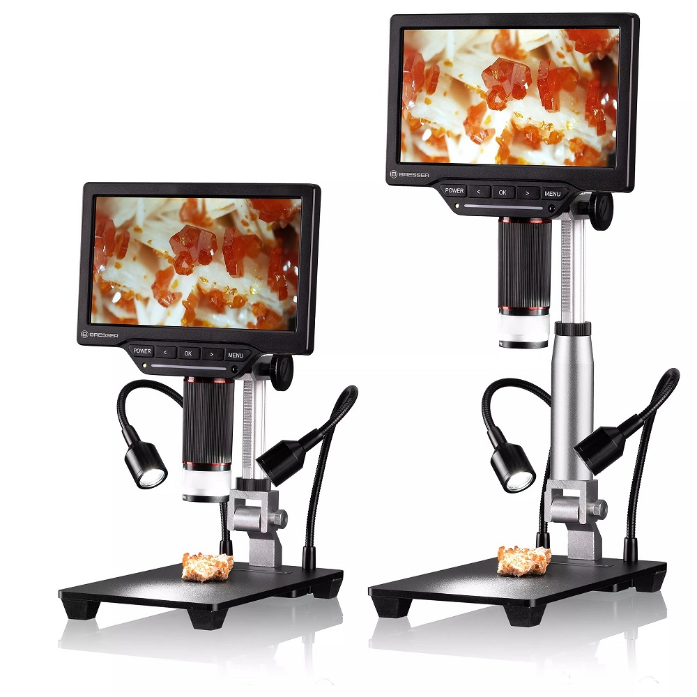
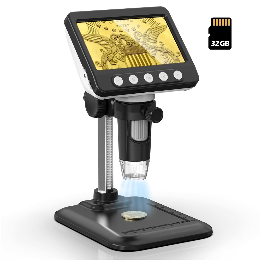
Digital microscopes come in various designs, each with specific features suited to distinct applications. Let’s explore the main types and their uses in educational and research settings.
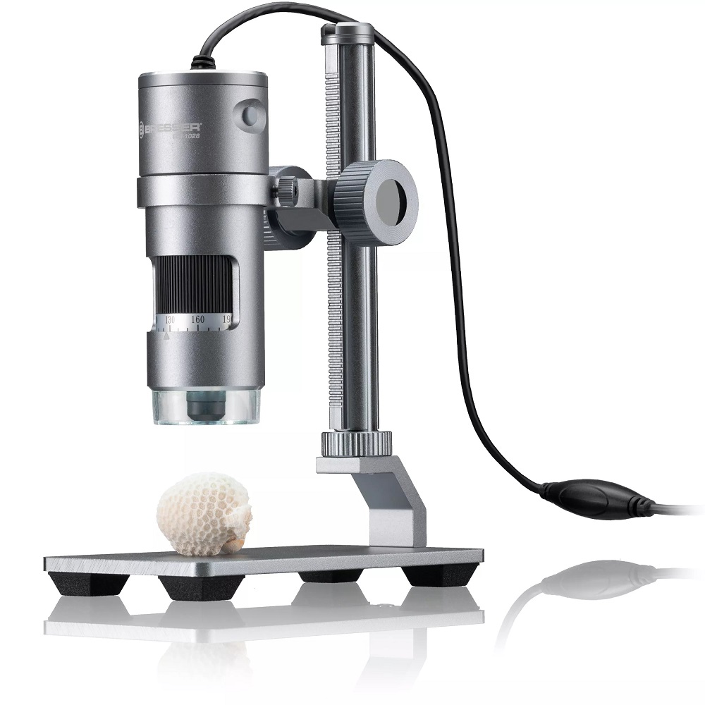
USB Digital Microscopes: These plug directly into a computer via USB. They are ideal for quick observations and for users who need to save and share images with ease.
Portable Digital Microscopes: Compact and battery-powered, these microscopes are perfect for fieldwork. Students can take them on trips to observe natural specimens in their habitats.
Wireless Digital Microscopes: These offer flexibility without the constraint of wires. They’re great for collaborative work, where the microscope can be passed around a classroom.
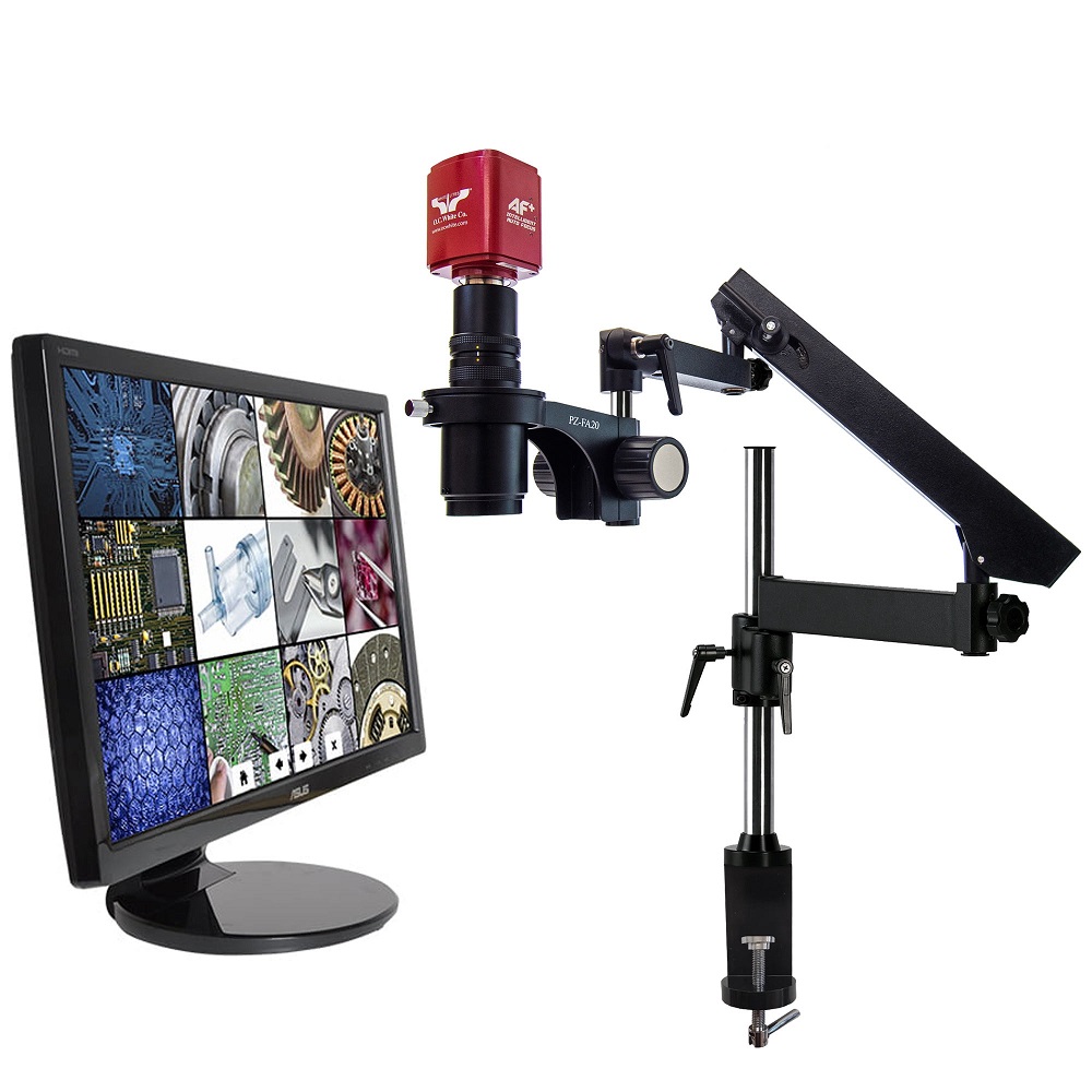
High-Resolution Digital Microscopes: These provide detailed images for advanced research. They are often used in higher education and professional labs.
Digital Microscopes with Measurement Software: Some models come equipped with software for measuring or analyzing specimens. This feature is particularly useful in material science and engineering education.
Each type of digital microscope has distinctive benefits that contribute to various aspects of learning and research. By choosing the right one, users can enhance their exploration of the micro-world. It’s important to match the microscope to the educational need or research demand to unlock its full potential.
Setting Up Your Digital Microscope for Optimal Results
To achieve the best possible outcomes when using your digital microscope, proper setup is key. Here are simple tips to ensure you’re getting the most out of your device:
Choose the Right Environment: Select a stable, well-lit area free from excessive movement and vibration. Good lighting enhances image clarity. A quiet setting reduces the chances of disruptions during sensitive observations.
Install Software Correctly: Before connecting your digital microscope to a computer, install any necessary drivers and software. Follow the instructions provided by the manufacturer to avoid technical issues.
Adjust the Focus: Start with a low magnification and slowly adjust the focus. Fine-tune it to get a sharp image.
Calibrate Your Device: If available, use calibration tools or software to ensure measurements and analyses are accurate. This step is crucial for any scientific work.
Secure the Specimen: Make sure the specimen is firmly placed on the stage. Movement can blur images and affect the quality of your observation.
Use Appropriate Lighting: Many digital microscopes have built-in illumination. Adjust the intensity so it’s neither too dim nor too harsh.
Check for Updates: Keeping your microscope’s firmware and software up to date can improve performance and introduce new features.
By following these simple steps, you’ll set yourself up for successful exploration with your digital microscope, enhancing both learning and research experiences.

Step-by-Step Guide to Operating a Digital Microscope
Operating a digital microscope is straightforward if you follow these steps.
Step 1: Set Up the Microscope
Begin by placing your digital microscope on a stable surface. Ensure it is connected to your computer or display.
Step 2: Install Necessary Software
Install any software that came with your microscope. This could be for image capture or analysis.
Step 3: Turn On the Device
Power on your digital microscope. Wait for the initialization process to complete.
Step 4: Place Your Specimen
Carefully position your specimen on the stage. Secure it to avoid movement.
Step 5: Adjust the Focus
Use the focus wheel to get a clear image. Start with low magnification and increase as needed.
Step 6: Set the Lighting
Modify the built-in lights for clear visibility. Avoid shadows and reflections on the specimen.
Step 7: Capture Images or Videos
With the subject in focus, capture images or record videos using the software.
Step 8: Analyze and Save
Use your digital microscope’s software to measure or analyze data. Save your findings.
Step 9: Clean Up
After use, clean the stage and lenses. Turn off and disconnect the microscope.
Remember, practice leads to perfect operation. Familiarize yourself with each step to maximize your digital microscope’s capabilities.

Common Pitfalls to Avoid When Using Digital Microscopes
As a professional blogger and SEO expert, I have gathered some tips to help you steer clear of common mistakes with digital microscopes. Here are pitfalls to avoid for a seamless experience:
Overlooking Calibration: Make sure to calibrate your digital microscope regularly. It ensures accuracy in measurements and image quality.
Ignoring Software Updates: Keep your software up-to-date. Outdated software can lead to compatibility issues and reduced functionality.
Neglecting Proper Lighting: Proper lighting is essential when using a digital microscope. Too much or too little light can spoil the clarity of your images.
Skipping Regular Maintenance: Clean the lenses and check the connections. Dust and dirt can affect the performance of your digital microscope.
Forgetting to Back Up Data: Always save and back up your images and videos. Losing data can be a significant setback in your research or education.
Using Incorrect Settings: Don’t rely on default settings. Adjust the magnification, focus, and brightness to suit your specific needs.
Rushing the Process: Take your time when focusing and capturing images. Rushing can result in blurred or low-quality results.
By avoiding these common errors, your work with a digital microscope will be more effective and rewarding. Remember, attention to detail and careful operation are key to unlocking the full potential of your digital microscope.
Enhancing Your Research with Digital Microscope Software Features
When diving into the capabilities of a digital microscope, software features play a crucial role. They can significantly enhance research outcomes. In this section, we’ll highlight how to maximize these features for superior results.
Advanced Image Analysis: Leverage tools that allow for detailed examination of captured images. Features like zoom, contrast, and brightness adjustments sharpen the details.
Measurement Functions: Use the measurement feature to determine the size and volume of specimens. Precise measurements are vital in many scientific studies.
Time-Lapse Recording: Record time-lapse videos to observe changes over time. This is great for documenting growth or decay in biological studies.
Annotation and Editing: Add notes and labels directly onto your images and videos. This helps with organizing data and sharing findings.
3D Reconstruction: Some software can create 3D models from multiple images. It provides a more comprehensive view of your subject.
Image Stacking: Combine several images taken at different focus points. It results in a final image with improved depth of field.
By tapping into these powerful software features, researchers can add value to their findings and gain deeper insights into their studies using the digital microscope.
Real-world Examples: Case Studies of Digital Microscope Usage
Digital microscopes are revolutionizing various fields through practical applications. Real-world case studies show their impact. Here are some noteworthy examples:
Educational Settings: A high school biology class used a digital microscope for a cell observation project. Students could view cells on a large screen, making learning dynamic and visually engaging.
Medical Laboratories: In a research facility, digital microscopes aided in the quick identification of pathogens. The ability to share images swiftly with colleagues sped up the diagnostic process.
Material Science: An engineering university utilized high-resolution digital microscopes. Students were able to examine material fractures in detail. This helped them understand stress points in different materials.
Environmental Studies: Scientists employed portable digital microscopes in the field. They documented new plant species in remote areas. The mobility of the devices made it possible to study specimens on-site.
Quality Control: A manufacturing company used digital microscopes with measurement software. They examined product microstructures for quality assurance. This reduced error rates and improved product reliability.
Forensic Science: Forensic experts used digital microscopes to analyze crime scene evidence. They could observe and record minute details that were crucial for investigations.
These case studies highlight just a few instances where digital microscopes advance knowledge and efficiency. They reveal how various sectors benefit from this technology. As digital microscopy evolves, its applications will further expand, opening new doors for discovery and innovation.
Future of Digital Microscopy: Trends and Advancements
The world of digital microscopy is evolving rapidly. Here we take a look at the latest trends and advancements that are shaping the future.
Integration with Artificial Intelligence (AI): AI is making digital microscopes smarter. With AI, microscopes can learn from past images and improve over time. They can identify patterns and anomalies faster. This leads to quicker and more accurate results.
Improved Image Quality: Advances in camera technology ensure even sharper images. High-resolution cameras capture intricate details. This is vital for complex scientific research.
Portability and Connectivity: New models are more portable and connect easily. They sync with various devices, improving convenience for users outside labs. This includes field researchers and educators on the go.
Automation: Automation in digital microscopes saves time. It allows for repetitive tasks, such as focusing and image capturing, to be done quickly. This frees researchers to focus on analysis and interpretation.
3D Imaging: Three-dimensional imaging features are becoming common. They allow for a more complete view of samples. This is especially useful in medical research and material sciences.
Cloud Integration: Cloud storage and computing are streamlining data sharing and collaboration. Data can be accessed anywhere, by multiple users, facilitating global research efforts.
Customization and Modularity: Users can customize digital microscopes to fit specific needs. Add-ons and modular designs let users upgrade their devices easily.
Virtual Reality (VR) Compatibility: VR can transform how we view microscopic images. It offers immersive educational experiences and could revolutionize scientific visualization.
As digital microscopy technology grows, researchers and educators will unlock new potentials. By staying up-to-date with trends, they will enhance learning and discovery. Schools and labs will be at the forefront of scientific progress.
