Introduction to Bacterial Microscopy
Bacterial microscopy offers us a window into a world unseen by the naked eye. This method lets scientists and researchers observe bacteria under microscope, revealing their complex structures and behaviors. The endeavor starts with choosing the right microscope. Various types yield different magnification levels. Light microscopes, for example, can magnify up to 1000 times. Electron microscopes go even further, offering an up-close look at bacteria. Staining is another step that enhances visibility. It colors different parts of the cells. This ease distinguishing between cell parts under high magnification. The knowledge gained from bacterial microscopy shapes our understanding of microbiology. It aids in research, diagnostics, and educational fields. As microscopes evolve, the clarity and detail in which we can view bacteria improve as well. The next sections will delve into the types of bacteria we study, how to prep samples, and microscopy techniques.
Types of Bacteria Commonly Studied Under the Microscope
When observing bacteria under microscope, certain types capture scientists’ focus frequently. These commonly studied bacteria include various species that affect human health, are fundamental in environmental ecosystems or hold industrial significance.
One prominent group is pathogenic bacteria, such as Staphylococcus aureus and Escherichia coli. These often cause diseases, hence the keen interest in their study. Understanding their structure helps in developing treatments.
Another group includes the beneficial bacteria from our gut, like Lactobacillus. These play a crucial role in digestion and overall health.
Environmental bacteria, such as the nitrogen-fixing Rhizobium, are critical for soil fertility.
Last but not least, industrial bacteria like Bacillus subtilis are used in fermentation processes. Their study is vital for optimizing production in industries.
These types give a glimpse of the diversity and impact of bacteria studied under microscopes. By magnifying these tiny organisms, we gain valuable insights into their world and ours.
Preparing a Bacterial Sample for Microscopic Examination
Before delving into the microscopic study of bacteria, proper sample preparation is crucial. It lays the foundation for clear and accurate observation. Here’s a step-by-step guide on how to get your bacterial sample ready for examination under a microscope.
First, obtain a culture of the bacteria you wish to study. This culture can come from a lab or an environmental sample. Ensure the culture is pure and uncontaminated for best results.
Next, you’ll need to transfer a small amount of the bacterial culture onto a microscope slide. This process is known as making a smear. Use a sterilized tool like a loop or needle for this and spread the bacteria thinly across the slide.
Once the smear is in place, it’s time to fix the bacteria to the slide. This is often done by gently heating the slide. This step prevents the bacteria from washing away during staining and allows for better adherence to the slide surface.
After fixing, staining the sample is the next step. Stains like crystal violet or methylene blue are common. They’ll help differentiate the bacteria from the background, making the structures more visible.
Finally, rinse off excess stain and allow the slide to dry. Once dry, it’s ready to be placed under the microscope. Using oil immersion can also enhance the resolution at high magnification. This technique involves placing a drop of oil between the slide and the lens to reduce light refraction.
By following these procedures for sample preparation, you can ensure that the bacteria under microscope will be displayed in the utmost clarity, ready for detailed examination and study.
The Different Microscopy Techniques for Observing Bacteria
To observe bacteria under microscope, several techniques are vital. These methods differ in their approach and the detail they reveal. The choice of technique often depends on the aspect of the bacteria being studied. Here is an overview of the primary microscopy techniques used for observing bacteria:
Light Microscopy: This is the most common method. It uses visible light to illuminate the sample. Simple stains often complement this technique to enhance contrast.
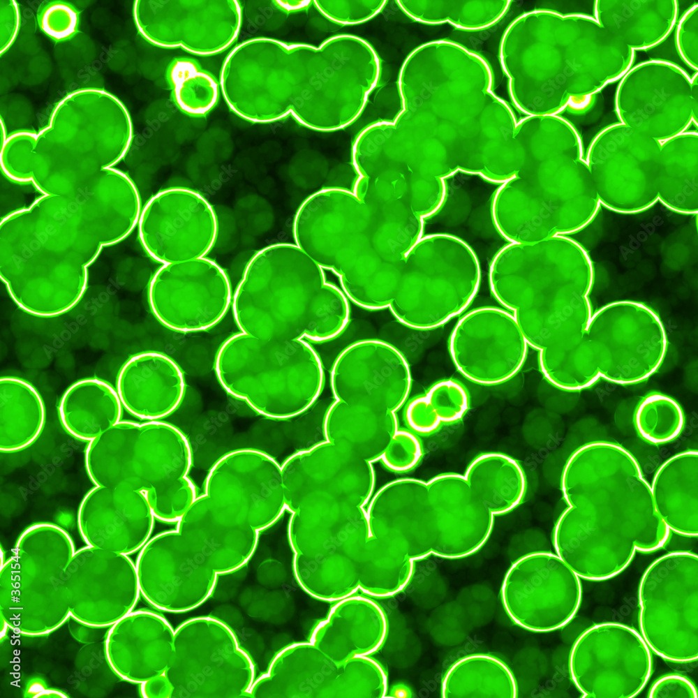
Fluorescence Microscopy: Here, fluorescent stains are used. They emit light when exposed to specific wavelengths. This method is great for tagging parts of the cell or specific proteins.
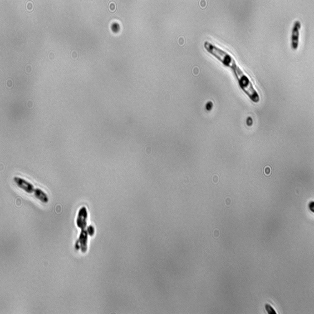
Phase-Contrast Microscopy: This technique enhances the contrast of transparent specimens without staining. It’s useful for live bacteria observation.
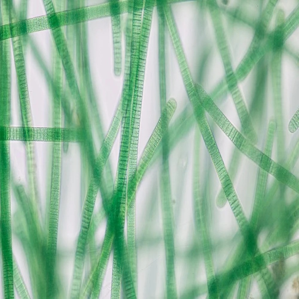
Dark-Field Microscopy: By blocking the central light, this method creates a contrast with a dark background. It’s excellent for visualizing spirochetes.
Electron Microscopy: There are two types:
- Transmission Electron Microscopy (TEM): Offers high-resolution images of bacteria structure. Thin sample sections are needed.
- Scanning Electron Microscopy (SEM): Shows the surface of bacteria in three dimensions. It provides a detailed topographical view.
Each technique serves a specific purpose and often, researchers may use multiple methods to get a comprehensive understanding. Choosing the right microscopy type can impact the level of detail observed in bacterial samples.
Understanding the Structure of Bacteria Through Magnification
Magnifying bacteria under a microscope transforms our perception of these tiny organisms. The structure of bacteria is complex, varied, and adapted to their environments and functions. Through magnification, we get a clear view of different bacterial components, such as cell walls, membranes, and nucleoids. Each component plays a role in the survival and behavior of bacteria.
Cell Walls and Membranes
Under high magnification, the bacterial cell wall is a prominent feature. It provides shape and protects the cell from external stress. When observing bacteria under microscope, one may notice that the cell wall’s appearance differs among species. Gram staining, a common technique in bacteriology, differentiates bacteria based on the structure of their cell walls.
Following the cell wall, the cell membrane is observed within. This membrane controls the movement of substances in and out of the cell. It is crucial for maintaining the life processes of the bacteria.
Cytoplasm and Internal Structures
The cytoplasm is the jelly-like substance filling the inside of the cell. Magnified views reveal various particles and structures within the cytoplasm. Ribosomes, responsible for protein synthesis, appear as small dots. Plasmids, small DNA molecules, may also be visible. They carry genes beneficial for bacteria, such as antibiotic resistance.
Nucleoid and Spores
Unlike higher organisms, bacteria do not have a defined nucleus. Instead, they have a nucleoid. The nucleoid is an irregularly shaped region that contains the bacterial DNA. Under a microscope, it is seen as a less dense area within the cytoplasm. Some bacteria can form spores, a survival mechanism under harsh conditions. These spores often stand out as distinct structures when magnified.
Magnifying bacteria reveals a lot about their structure and behavior. Techniques like Gram staining, along with advanced microscopy methods, allow scientists and researchers to identify and understand bacteria. This enhanced knowledge is important for various applications, including medical treatments and biotechnology.
Microscopic Photography: Capturing Bacteria in Detail
Microscopic photography has revolutionized how we view bacteria under microscope. It involves capturing detailed images of bacteria for further study. This is critical for research, diagnostics, and education. These high-resolution images can show tiny details. Colors become clearer, and shapes become more defined against the magnified backdrop. For capturing these details, photographers often rely on digital cameras attached to microscopes. Such setups allow for instant image viewing, editing, and sharing.
Techniques in Microscopic Photography
To get the best photographs, different methods come into play. Focus stacking is a technique where multiple images at different focal lengths are combined. This creates a single image with greater depth of field. Another method is differential interference contrast (DIC) microscopy. DIC gives a three-dimensional effect to flat specimens. It enhances the texture and details.
Challenges in Capturing Images of Bacteria
Photographing bacteria presents unique challenges. Bacteria can move quickly, making it tough to get a clear shot. The high magnification required can also lead to issues with depth of field and lighting. Specialized equipment and techniques, like high-speed cameras and proper staining, help address these challenges. Careful sample preparation is also key. It ensures the bacteria are still and well-defined when photographed.
Microscopic photography demands skill and patience but offers invaluable outputs. This is not just for the scientific community, but also for public understanding. Photographers can weave stories through the images. They can showcase the hidden beauty and complexity of bacteria under microscope. These photos are more than just science; they’re works of art that reveal an unseen world.
The Role of Microscopes in Bacterial Research and Diagnostics
Microscopes play a crucial part in studying bacteria. They help us see bacteria structures we can’t with our eyes. In research, scientists use them to learn how bacteria live and grow. This understanding leads to new ways to fight infections. Doctors also use microscopes to spot harmful bacteria in patient samples. This is key to diagnosing diseases quickly and accurately.
Microscopes have become vital tools in tracking disease outbreaks too. Public health experts use them to identify bacteria strains in an outbreak. This helps in controlling the spread of disease. Advanced microscopes can even show how bacteria react to antibiotics. This is important in developing resistance strategies.
Microscopes are not just for professionals. They are used in classrooms to teach about bacteria. Students can see first-hand the tiny organisms they learn about. This makes learning about bacteria exciting and real.
In sum, microscopes are essential in the fight against bacterial diseases. They are important tools in research, diagnostics, and education. They help us see and understand the unseen world of bacteria under microscope.
Advances in Microscopic Technology for Bacterial Study
The field of bacterial study has seen significant advances in microscopic technology. These advances have made observing bacteria under microscope more detailed and precise than ever. One major development is in high-throughput microscopy. This allows researchers to study many samples at once, saving time and resources. New imaging techniques now offer higher resolution. They permit scientists to view bacterial processes in real time.
Digital microscopy is another key advancement. It combines traditional microscopy with digital tech. Users can now store images and analyze them with software. This makes identifying and tracking bacteria much easier. Automation in microscopes adds to this efficiency. It reduces the need for manual adjustments during study.
Super-resolution microscopy breaks the limits of traditional light microscopy. It lets us see structures within bacteria that were too small before. This type of tech is crucial for understanding bacterial function at molecular levels.
Live-cell imaging has also improved. It involves watching bacteria as they grow and interact. This gives insights into their behavior in environments similar to their natural habitats.
In sum, these technological improvements allow for deeper investigation. We can explore the intricate world of bacteria under microscope in ways not possible before. They push the boundaries of what researchers can discover about bacterial life.