
Introduction to Blood Cell Microscopy
The study of blood under microscope unveils a world teeming with vital cellular activities. Blood cell microscopy is a critical laboratory technique. It allows scientists and medical professionals to observe and analyze the characteristics of blood cells. This analysis aids in understanding not only the health of the cells themselves. It also provides insights into the overall well-being of an individual.
Using powerful microscopes, experts can detect and scrutinize various blood cell types, structures, and any potential abnormalities present. This microscopic examination is an essential step in many medical diagnostics. It is the foundation for treating diseases and monitoring patient health.
Blood cell microscopy requires precision and expertise. Equipment used includes traditional light microscopes, and more advanced tools like electron microscopes. These instruments magnify blood samples significantly. They reveal details that are imperceptible to the naked eye.
Overall, blood cell microscopy stands as a cornerstone in medical science. It offers a window into the complex cellular composition of our blood. It plays a fundamental role in the diagnosis and treatment of countless conditions. Researchers and healthcare providers rely on this technology to deliver accurate medical care.

Types of Blood Cells and Their Functions
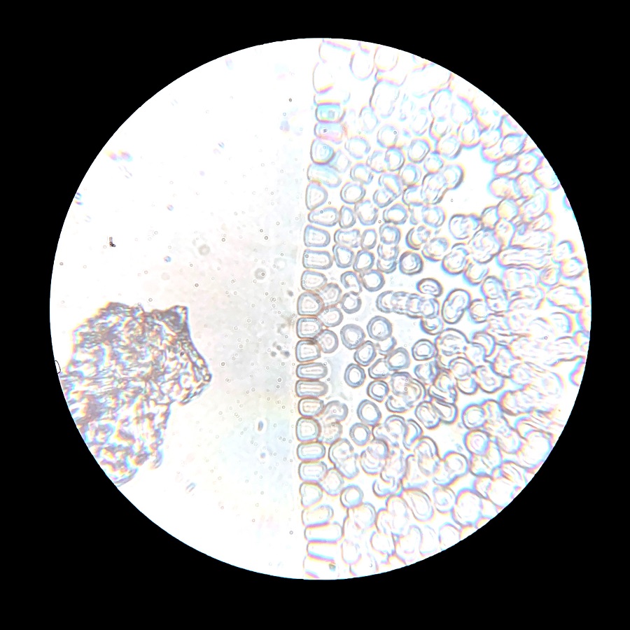
Examining blood under microscope reveals three primary types of blood cells. These are red blood cells (RBCs), white blood cells (WBCs), and platelets. Each type has unique features and critical functions in the body.
Red Blood Cells (Erythrocytes)
Red blood cells are the most abundant. They carry oxygen from the lungs to the body’s tissues. They also transport carbon dioxide back to the lungs for exhalation. Their biconcave shape maximizes the cell’s surface area. This shape enhances their oxygen-carrying capacity.
White Blood Cells (Leukocytes)
White blood cells are key players in the immune system. They protect the body against infections and foreign invaders. There are several types of white blood cells. These include lymphocytes, neutrophils, eosinophils, basophils, and monocytes. Each type has a specific role in defense mechanisms.
Platelets (Thrombocytes)
Platelets are tiny cells that play a critical role in blood clotting. They prevent excessive bleeding when injuries occur. Upon vessel damage, platelets clump together and release chemicals. These chemicals initiate the clotting process.
Understanding the functions of these blood cells is vital. With blood under microscope, medical professionals can assess an individual’s health. They track the functioning of each blood cell type. When blood cell counts or appearances deviate from the norm, it may indicate health issues. Thus, blood cell microscopy is crucial in routine health check-ups and responding to diseases.
Preparing a Blood Sample for Microscopic Examination
Before placing blood under microscope, proper preparation is key. This ensures accurate and detailed observations. Here’s a step-by-step guide on preparing a blood sample for microscopic examination:
- Sample Collection: Blood is typically drawn from a vein using a sterile needle. This process, known as venipuncture, must be carried out by a trained professional.
- Anticoagulants: Immediately after collection, blood is mixed with anticoagulants. This prevents clotting, which could distort the microscopic analysis.
- Sample Dilution: In some cases, the blood sample may be diluted. This helps distinguish individual cells more clearly when they are too numerous.
- Staining: Stains are often used to enhance visibility of blood cell components. For instance, Wright’s stain differentiates cells, making them easier to identify and count.
- Smear Preparation: A small drop of blood is spread thinly across a slide to create a ‘smear’. This thin layer allows light to pass through and cells to be examined individually.
- Fixing the Smear: The blood smear is usually air-dried and then fixed. Fixation preserves cell structure and prepares the sample for staining.
- Cover Slipping: Finally, a cover slip is placed over the smear. This protects the sample and eliminates air bubbles that could interfere with microscope lenses.
By following these steps, professionals prepare blood for the meticulous analysis under microscope. It’s a process that requires attention to detail and precise handling of the blood sample. Once prepared, the slide is ready for the microscope. Here, experts will scrutinize the sample for a detailed assessment of blood cell health and functioning.
The Process of Blood Cell Analysis Under the Microscope
When examining blood under a microscope, there is a structured process that professionals follow for accurate analysis. Once the blood sample is prepared and placed on a slide, the in-depth examination begins.
- Microscope Calibration: Before starting, the microscope gets adjusted to ensure clear images.
- Scanning the Slide: The slide is scanned at a lower magnification to locate areas rich in cells.
- Selecting the Field of View: A higher magnification focuses on selected regions with diverse cell types.
- Observing Cell Morphology: Specialists observe cell shape, size, and structure, noting any peculiarities.
- Counting Blood Cells: Red blood cells, white blood cells, and platelets are counted. This helps measure their concentrations in the blood.
- Identifying Cell Types: Different types of white blood cells are identified by their size, shape, and staining patterns.
- Noting Abnormalities: Any irregular cells, like those that are misshapen or unusually sized, are marked for further analysis.
- Documenting Findings: Observations and counts are recorded in detail for diagnosis and treatment plans.
This analysis is crucial for understanding the state of a patient’s health. Changes in blood cell numbers or structures often signal health issues. By examining blood under microscope, early detection of diseases is possible, aiding in timely treatment and better outcomes.
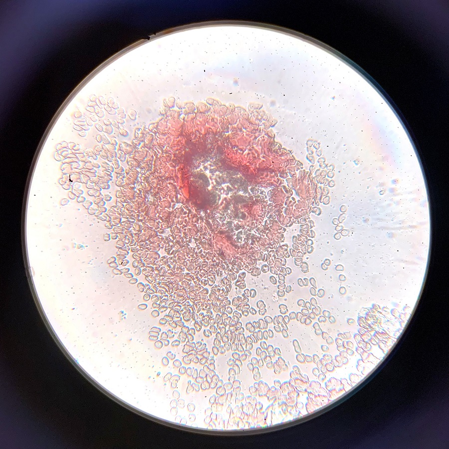
Common Blood Cell Abnormalities and Their Indicators
Identifying blood cell abnormalities is crucial when examining blood under microscope. Certain indicators can alert experts to potential health issues that may require further investigation. Below are some common irregularities and what they might suggest about a person’s health.
Red Blood Cell Abnormalities
Abnormalities in red blood cells (RBCs) often show up as changes in size or shape. For instance, when RBCs are larger than normal, a condition known as macrocytosis, it might indicate vitamin B12 or folate deficiency. Smaller RBCs, a condition called microcytosis, may suggest iron deficiency anemia. Misshapen RBCs can point to a range of disorders, including sickle cell anemia or hereditary spherocytosis.
White Blood Cell Abnormalities
White blood cells (WBCs) fight infection and react to stress in the body. When WBC counts are high, it typically signals infection, inflammation, or even leukemia. Low WBC counts may suggest bone marrow problems or autoimmune conditions. Observing the shape and structure of WBCs can also provide clues; abnormal appearances might indicate leukemia or other serious conditions.
Platelet Abnormalities
Platelets are essential for clotting. Too many platelets, a condition called thrombocytosis, can increase the risk of clots. Too few platelets, known as thrombocytopenia, may lead to excessive bleeding. Irregular platelet function can also be an indicator of various hematologic diseases.
By understanding these abnormalities and their potential health implications, medical professionals can make better diagnostic decisions. With ongoing advances in technology, blood under microscope serves as a powerful tool for early detection and treatment of many conditions.
Innovations in Blood Microscopy Technology
Recent advancements in blood microscopy technology have greatly enhanced our ability to examine blood under microscope. These innovations lead to more accurate diagnoses and better patient care. Some notable developments include:
- Digital Microscopy: Unlike traditional microscopes, digital systems capture and store images digitally. This allows for easier sharing and analysis of blood cell images.
- Automated Cell Counters: These devices automate the counting of blood cells, increasing the speed and accuracy of the process.
- High-Throughput Screening: Newer technologies enable the examination of thousands of blood samples quickly. This is essential for large-scale studies or screenings.
- Fluorescence Microscopy: Using special dyes that glow under certain light, this method can highlight specific blood cell components or abnormalities.
- Live Cell Imaging: Advancements now allow for the observation of live blood cells in action. This helps in understanding dynamic processes like clot formation.
- AI Integration: Artificial intelligence (AI) is being used to detect patterns and predict conditions based on blood cell images.
Each innovation brings us closer to real-time, accurate blood analysis. Doctors and researchers can now uncover details about blood cells and their health with unprecedented clarity. These strides in technology pave the way for better detection and treatment of diseases. Given these tools, the future of blood cell microscopy seems bright, with potential for even more groundbreaking discoveries.
The Role of Blood Cell Microscopy in Medical Diagnoses
Blood cell microscopy is crucial in medical diagnoses. It allows doctors to make informed decisions based on cellular evidence. When health issues arise, a clear understanding of blood cells matters greatly. Medical professionals turn to microscopy to look at blood under microscope. This process is integral to pinpointing diseases and planning treatments.
Here are key reasons why blood cell microscopy is vital in diagnoses:
- Early Disease Detection: Examining blood cells can reveal early signs of conditions. Early signs such as anemia, infections, or clotting disorders are visible when blood is under microscope. This means treatments can start sooner, often leading to better outcomes.
- Understanding Infection Severity: High white blood cell counts can indicate serious infections. Microscopy helps assess how severe an infection is and guides doctors on the best course of action.
- Monitoring Treatment Progress: Patients receiving treatment for diseases like cancer often undergo blood tests. Doctors use blood cell microscopy to monitor how well a patient responds to treatment. Changes in blood cell health can indicate improvement or the need for a different approach.
- Personalized Medicine: Every patient’s blood tells a unique story. By examining blood under microscope, doctors can tailor treatments to the individual needs of each patient.
- Implementing Preventive Measures: Regular checks of blood cell health can prevent diseases. For instance, spotting slight changes in blood cell health can prompt lifestyle adjustments or early treatment.
In summary, blood cell microscopy is not just a tool for diagnoses. It is a constant companion in managing patient health. It serves as eyes for doctors, revealing what might otherwise remain unseen. By understanding blood cells in minute detail, medicine continues to advance and save lives.
Future Directions in Blood Cell Microscopic Research
Exploring the future of blood cell microscopic research, scientists are heading towards remarkable territories.
With ongoing technological leaps, researchers foresee a transformation in how we examine blood under microscope. Future advancements may include:
- Enhanced Imaging Techniques: Nanotechnology and improved digital imaging could provide even more detailed views of blood cells.
- Microfluidics Integration: This technology allows precise manipulation of tiny blood volumes, enhancing sample analysis.
- Portable Devices: The development of handheld microscopes could make blood analysis accessible in remote areas.
- Cloud-based Analysis: Storing and analyzing blood cell images in the cloud may enable real-time collaboration among global experts.
- Gene Editing: CRISPR and other gene-editing tools might allow researchers to study blood cells directly from the genetic level.
- Personal Health Monitoring: Wearable technology could one day track blood cell health, offering personalized medical insights.
- Machine Learning Algorithms: Advanced AI could be trained to recognize complex patterns in blood cells, speeding up diagnoses.
The focus of blood cell microscopic research is shifting. It aims to not just observe, but also interact with and modify cells at an unprecedented scale. When considering blood under microscope, these innovations promise to expand our knowledge. They may lead to groundbreaking treatments and a deeper understanding of human biology.