Introduction to Plant Cell Microscopy
Delving into the microscopic world reveals a fascinating reality about plant cells. Observing a plant cell under a microscope can unearth its complex architecture and vivid processes. Microscopy has been a critical tool for scientists looking to understand the structure and function of plant cells. It allows us to observe cells in great detail, including the tiny organelles that are the building blocks of plant life.
For those new to botany or biology, plant cell microscopy is a revealing first step. It shows us how plant cells differ from animal cells, and how they work to sustain the plant organism. When we place a plant tissue slice under the microscope, we enter a world of cell walls, chloroplasts, and nuclei. These structures are essential to plant life, dealing with tasks such as photosynthesis, nutrient transport, and reproduction.
To get started with plant cell microscopy, one needs a good-quality microscope. It should have the ability to magnify cells sufficiently to see their components. We also need to prepare slides correctly, staining them if needed, to enhance the visibility of different cell parts. The goal is a clear image that shows us the wonders of the cell. A microscope with advanced features can show more details, but even a basic model can provide exciting insights.
In upcoming sections, we’ll discuss how to prepare plant tissues for examination, types of microscopes suited for this purpose, and the specific structures to look out for. Keep in mind, as we explore the cellular level of plants, that every vivid image and every new discovery has wide-ranging implications. From agriculture to medicine, the study of plant cells under a microscope informs many fields, driving innovation and understanding of the natural world.

Preparing Plant Tissues for Microscopic Examination
To study a plant cell under a microscope, proper tissue preparation is crucial. The process begins with selecting healthy plant tissue, which ensures that the cells observed are in good condition. After selecting the tissue, it is then carefully sliced into thin sections. These sections, or slices, must be thin enough to allow light to pass through when placed under the microscope.
Next, the thin plant slices are typically treated with a stain. This stain helps to contrast different parts of the cell, making it easier to see specific structures within the plant cell. For many stains, a waiting period is necessary to allow the dye to penetrate the cell components. After staining, the tissue is gently rinsed to remove any excess stain. Finally, the stained tissue is placed onto a glass slide and secured with a cover slip. This step prevents air bubbles and keeps the tissue flat for optimal viewing.
Throughout the preparation process, careful handling is essential to maintain the integrity of the plant specimen. It’s also helpful to label the slide with the plant’s name and the stain used, for accurate record-keeping and reference. Following these steps ensures that the plant cells are ready for examination and will provide a clearer understanding when viewed through a microscope lens.
Microscope Types and Techniques for Viewing Plant Cells
When exploring the structure of a plant cell under a microscope, the type of microscope used is vital. Different microscopes provide varying levels of magnification and resolution, which can influence the detail and clarity of the cellular image observed. Here, we will look at the common types of microscopes and techniques used for viewing plant cells.
Light Microscopy
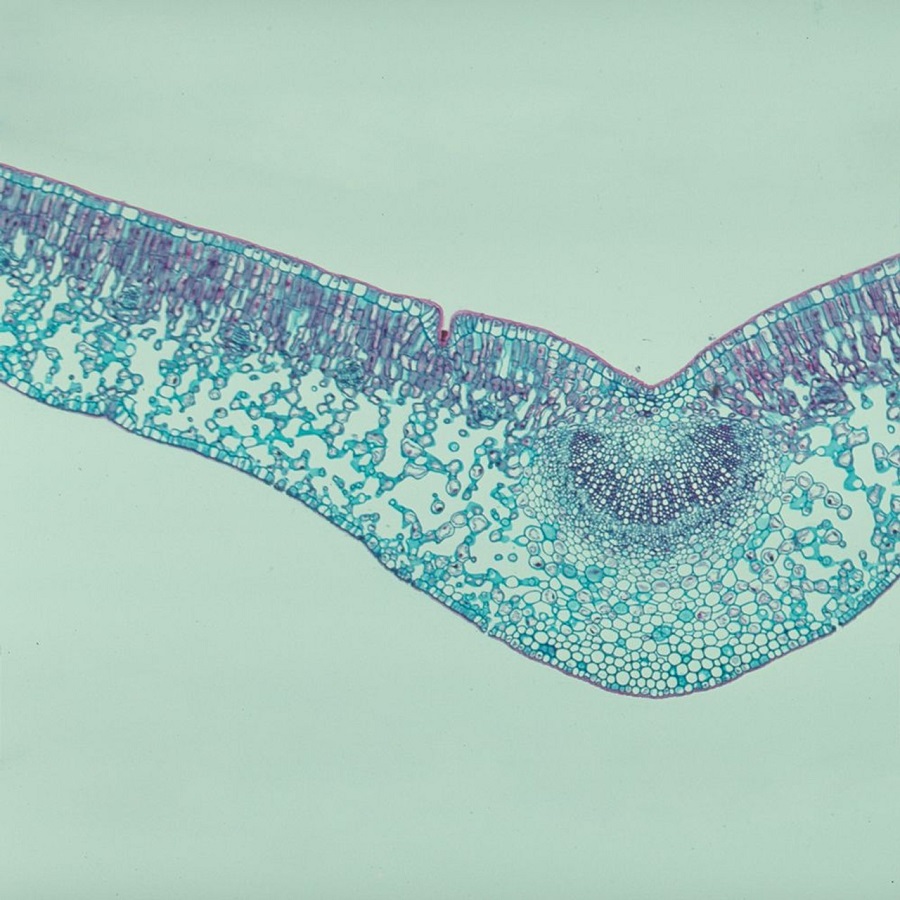
The most accessible type of microscope for beginners is the light microscope. It uses visible light to illuminate the sample and can magnify cells up to 1000 times. Preparing plant tissues involves staining, as discussed previously, which is especially effective with light microscopy to highlight cellular structures.
Electron Microscopy
For a more in-depth examination, electron microscopes are used. These offer much greater magnification and resolution, allowing scientists to see details at the molecular level. There are two main types – transmission electron microscopes (TEM) and scanning electron microscopes (SEM). TEM provides detailed internal cell views, whereas SEM offers a three-dimensional look at the cell’s surface.
Fluorescence Microscopy
This technique involves using fluorescent stains that emit light when excited by specific wavelengths. Fluorescence microscopes can thus reveal specific structures within plant cells, such as proteins or DNA, by using different fluorescent compounds.
Confocal Microscopy
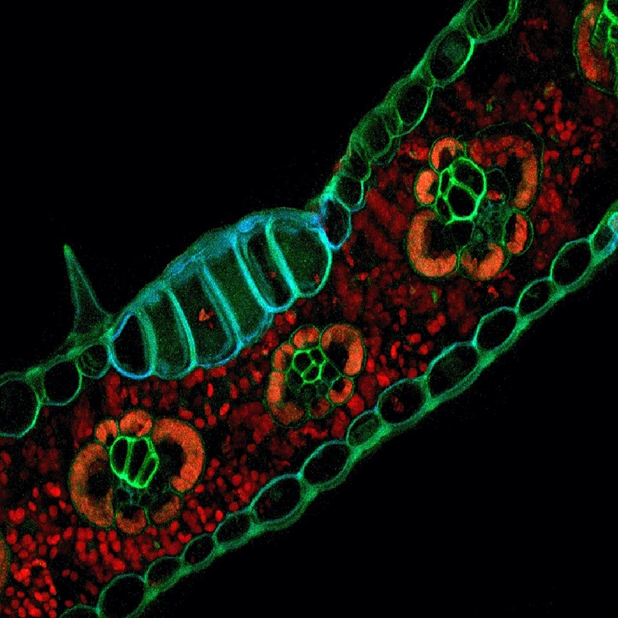
Confocal microscopy enhances optical resolution and contrast by using a spatial pinhole to block out-of-focus light. It allows for the construction of three-dimensional images from the collected data and is particularly helpful in detailed studies of plant cell structures.
Phase Contrast and Differential Interference Contrast (DIC) Microscopy
Both phase contrast and DIC microscopes are designed to enhance contrast in transparent specimens without the use of stains. These are useful for live cell imaging, allowing scientists to observe plant cells in their natural state.
In summary, understanding which microscope and technique to utilize is key to effectively examining a plant cell under a microscope. Each method has its own strengths and is chosen based on the specific requirements of the study at hand. Most importantly, using the correct technique illuminates the intricate details of plant cells that are not visible to the naked eye.

The Fundamentals of Plant Cell Structure
When a plant cell is placed under a microscope, a whole new world of structures opens up. Each cell is like a busy city, with various organelles performing vital functions. Understanding the basics of their structure helps us appreciate how plants thrive. Here are the key components of a plant cell:
- Cell Wall: Unlike animal cells, plant cells have a rigid cell wall. This wall provides structural support and protection. It’s made mostly of cellulose.
- Plasma Membrane: Just inside the cell wall is the plasma membrane. It controls the movement of substances in and out of the cell.
- Cytoplasm: The cytoplasm is a jelly-like substance where many cell processes occur. It holds the organelles in place.
- Nucleus: Acting as the control center, the nucleus contains DNA. It regulates cell growth and reproduction.
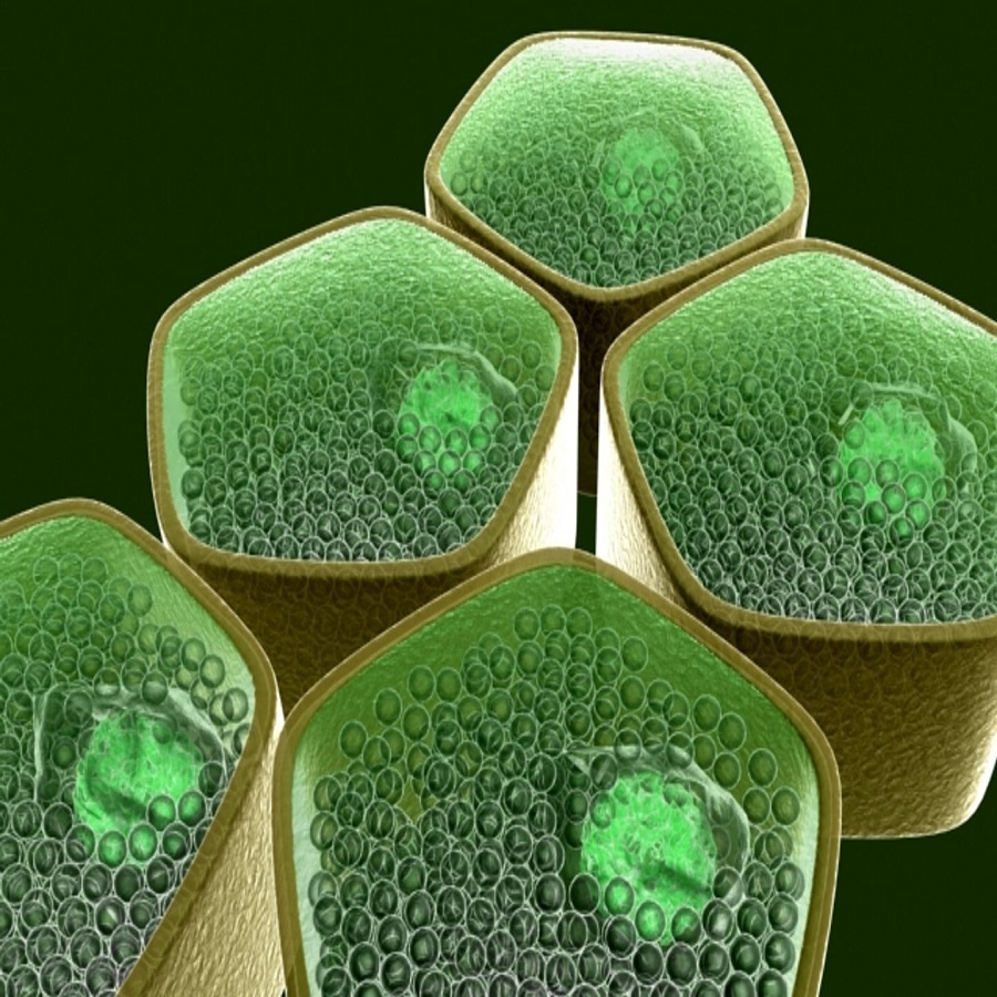
- Chloroplasts: These are the sites of photosynthesis. Chloroplasts capture sunlight and use it to produce food for the cell.
- Mitochondria: Known as the powerhouses, mitochondria generate the cell’s energy. They convert glucose into ATP, which powers cellular activities.
- Endoplasmic Reticulum (ER): The ER comes in two forms – rough and smooth. It’s involved in protein and lipid synthesis.
- Golgi Apparatus: This structure modifies, sorts, and packages proteins for delivery throughout the cell.
- Vacuoles: Plant cells typically contain a large central vacuole. It stores nutrients and waste products and also maintains cell pressure.
- Lysosomes and Peroxisomes: These organelles break down waste materials and toxins within the cell.
Each component plays an intricate part in the cell’s survival and function. Through the lens of a microscope, we can observe these structures in detail. This observation can lead to a deeper understanding of plant biology and the vital roles that plant cells fulfill in our ecosystem.
Observing Chloroplasts and Photosynthesis Process
When we place a plant cell under a microscope, one of the most striking features we might see are the chloroplasts. These green structures are the centers of photosynthesis, the process through which plants convert sunlight into chemical energy. Let’s delve into how chloroplasts appear under the microscope and their role in the photosynthesis process.
- Appearance of Chloroplasts: Under the microscope, chloroplasts are typically easy to spot due to their green pigment. They are oval-shaped and may appear to move within the cell, a phenomenon known as cytoplasmic streaming.
- Photosynthesis in Action: While the process of photosynthesis is not visible, the chloroplasts are where it happens. They contain chlorophyll, which captures sunlight. Through a series of reactions, the cell turns this energy into glucose, a source of food.
- Staining Chloroplasts: To observe chloroplasts more clearly, biologists might use specific stains. These can highlight chloroplasts, making them stand out against the rest of the cell structures.
- Observing the Process: During the peak photosynthetic activity, you can sometimes see an increase in movement within the chloroplasts under the microscope. This indicates the active processes happening within.
Viewing chloroplasts under the microscope offers insight into the incredible energy conversion plants perform every day. It’s a critical step in understanding how plants sustain themselves and support life on Earth.
Identifying Organelles Unique to Plant Cells
When studying a plant cell under a microscope, specific organelles unique to plant cells stand out. These structures distinguish plant cells from animal cells and are crucial for various plant functions.
- Chloroplasts: Unique to plants and some algae, chloroplasts perform photosynthesis. Visible under a microscope, they are green due to chlorophyll.
- Cell Wall: The cell wall’s rigid structure is observable around the plant cell’s perimeter. It provides strength and support.
- Central Vacuole: Occupying a significant portion of the cell’s volume, the large central vacuole can often be seen. It’s essential for maintaining turgor pressure.
- Plasmodesmata: Not visible with all microscopes, plasmodesmata are channels within the cell wall, allowing transport between cells.
- Leucoplasts and Chromoplasts: These are plastids involved in storage and pigment synthesis, respectively. They may not be as prominent as chloroplasts.
Identifying these features helps illuminate the intricate network of plant cell operations. Recognizing them enhances our understanding of their key roles in plant vitality and survival.

Comparison Between Plant and Animal Cells Under the Microscope
When examining a plant cell under microscope, we notice distinctive differences from animal cells. Here’s a comparison of some features you might observe:
- Cell Walls: Plant cells have a rigid cell wall made of cellulose, which animal cells lack.
- Chloroplasts: These are present in plant cells but not in animal cells, signifying their role in photosynthesis.
- Vacuoles: Plant cells often contain a large central vacuole. Animal cells may have small vacuoles, but typically not as prominent.
- Shape: Plant cells usually have a regular, rectangular shape, while animal cells have varied and less defined shapes.
- Lysosomes: These are more common in animal cells, whereas plant cells have fewer lysosomes and rely more on peroxisomes.
- Centrioles: These are found in animal cells aiding in cell division but are absent in most plant cells.
This comparison not only helps distinguish plant cells from animal cells but also highlights their unique functions. It allows us to understand how cells are adapted to their roles in different organisms.
Advanced Imaging Techniques and Their Applications in Plant Cell Research
Advancements in imaging technology have enriched our knowledge of plant cell biology. Here we discuss the role of advanced imaging techniques and their applications in plant cell research.
- Super-Resolution Microscopy: This technology surpasses the limits of traditional light microscopy, enabling scientists to view structures at the nanoscale. Researchers can now see details of organelles previously blurred.
- Multiphoton Microscopy: In this technique, cells absorb two photons simultaneously, allowing for deeper tissue penetration. It is ideal for viewing live tissue without causing damage.
- Atomic Force Microscopy (AFM): AFM maps the surface of cells at an atomic level. With AFM, scientists examine the physical properties of plant cell walls.
- Total Internal Reflection Fluorescence (TIRF) Microscopy: This method provides high-resolution images of cell surface events. It’s helpful for studying how cells interact with their environment.
- Live Cell Imaging: Tracking cellular processes in real-time is possible with live cell imaging. It lets researchers observe how cells grow, divide, and respond to stimuli.
Each technique offers unique insights. For example, super-resolution microscopy has been critical for understanding how proteins organize within cells. Multiphoton microscopy has aided in studying plant cell interaction with pathogens.
These techniques have applications beyond basic research. They contribute to agriculture by helping scientists design crops more resistant to disease and environmental stress. In medicine, understanding plant cells aids in developing plant-derived drugs.
The advanced imaging methods transform the scope of plant cell study. They empower researchers to uncover minute details that can have significant impacts on our understanding of plant biology and its applications.
The Role of Microscopy in Plant Biology Education
Microscopy has a crucial part in teaching plant biology. By using microscopes, students gain a direct view of what they learn about in textbooks, deepening their understanding of plant cell structure and function. Here are key ways in which microscopy is integral to plant biology education:
- Hands-On Learning: Microscopy provides a hands-on approach. Students can see the cells and organelles they’re studying, which makes the learning experience more engaging and tangible.
- Enhanced Comprehension: Visualizing a real plant cell under microscope helps students grasp complex concepts. It’s one thing to read about chloroplasts, but seeing them brings lessons to life.
- Discovery and Inquiry: Students can explore plant cells and ask questions based on their observations. This inquiry-based learning fosters critical thinking and scientific curiosity.
- Practical Skills Development: Working with microscopes helps students develop practical skills. They learn how to prepare slides, handle equipment, and document their findings.
- Inspiring Future Scientists: By observing the wonders of plant cells up close, students might be inspired to pursue careers in botany or related scientific fields. Microscopy can light up paths to such opportunities.
In summary, microscopes are not just tools; they are bridges to a better understanding of plant biology. They inspire and educate, making learning an active and exciting process. Every time students look at a plant cell under microscope, they’re not just seeing a cell; they’re getting a glimpse of the complexity and beauty of life itself.