Microscopes are essential tools in various scientific fields, from biology to materials science. To effectively utilize a microscope, it’s crucial to understand its components. This comprehensive guide will explore the parts of microscope labeled, providing you with detailed insights into each part’s function and importance. Whether you’re a student, educator, or professional, knowing the labeled parts of a microscope will enhance your ability to conduct precise and efficient observations.
 Introduction to Microscope Structures
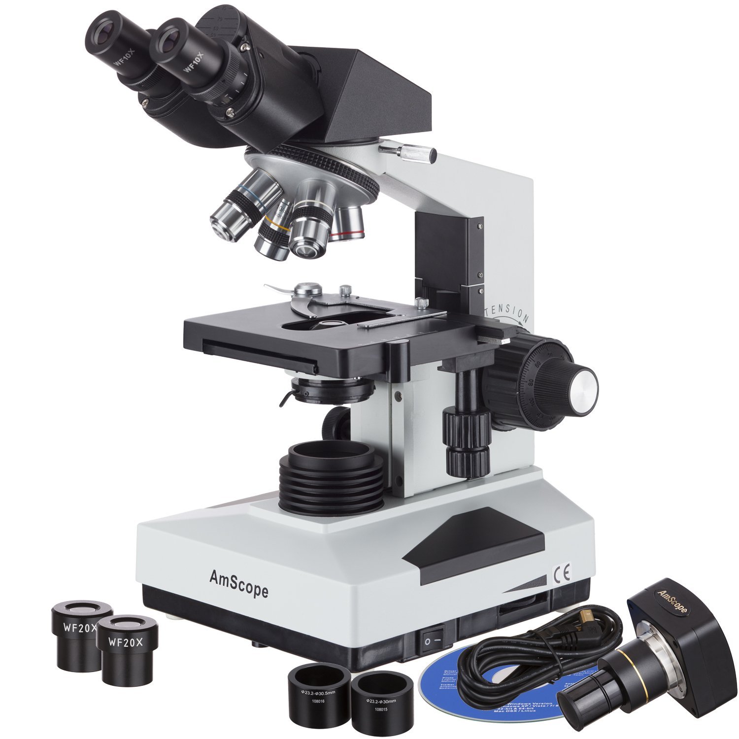
Microscopes are vital tools in many scientific fields. They consist of various parts that each play a crucial role in magnifying small objects. Understanding the distinct parts of a microscope helps users operate them effectively. Let’s explore these components.
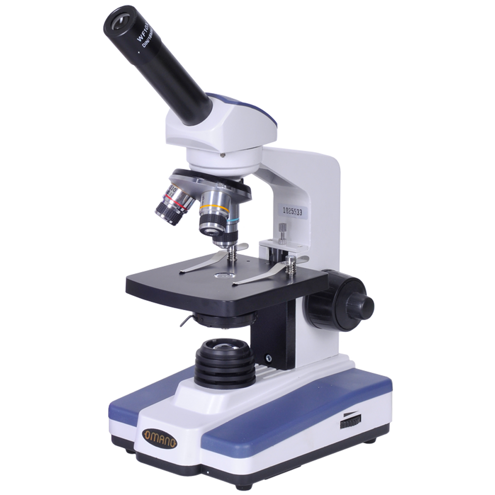
Eyepiece or Ocular Lens
This is where you look through to see the magnified image of the sample. The eyepiece usually contains a lens that enhances the magnification of the specimen.
Objective Lenses
Found on the nosepiece, these lenses are the primary magnifiers of the microscope. They come in different powers, such as 4x, 10x, 40x, and sometimes even 100x.
Stage
The platform where the slides holding samples are placed. It often comes with clips to secure the slides in place.
Light Source
Located beneath the stage, it illuminates the specimen, making details visible and easier to study.
Coarse and Fine Focus Knobs
These knobs adjust the focus of the microscope to sharpen the image. The coarse focus is used for general focusing, while the fine focus is for detailed refinement.
Diaphragm or Condenser
Regulates the amount of light that reaches the specimen. Adjusting the diaphragm improves contrast and detail in the observed object.
These labeled parts of the microscope work in harmony to provide a clear, magnified view of microscopic worlds. As you delve deeper into each component’s functionality, you’ll appreciate the intricate design of these scientific instruments. The ‘parts of microscope labeled’ enhance understanding, enabling users to get the most out of their microscopy experiences.
 Key Components of a Microscope
A microscope is composed of several key parts that enable it to function effectively. Each component has a specific role that contributes to the overall operation of the microscope. For those who are using microscopes, whether in educational settings, research labs, or for hobbyist purposes, familiarizing yourself with these parts is crucial.
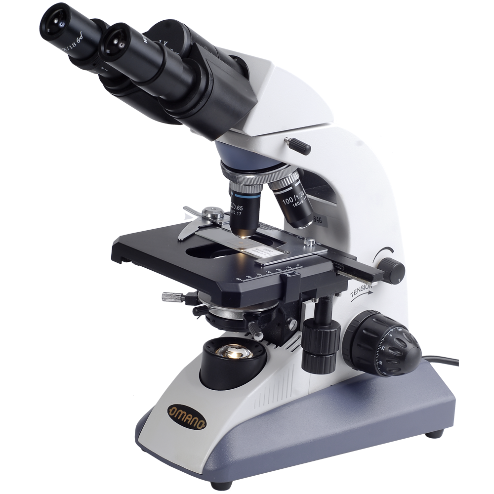
Base and Arm
The base provides stability, holding the microscope steady during use. It is the bottom support of the microscope. Attached to the base, the arm is what you grasp to carry the microscope safely.
Revolving Nosepiece
The revolving nosepiece, or turret, holds the objective lenses and can be turned to change magnification levels easily.
Mechanical Stage
An advanced feature on some microscopes, the mechanical stage allows precise movement of the slide in both the X and Y axes for detailed examination.
Illuminator and Mirror
The illuminator is an in-built light source that directs light upwards onto the slide. In some models without an electric illuminator, a mirror is used to reflect ambient light.
Adjustment Knobs
The adjustment knobs include both the coarse and fine focus knobs. They control the focus of the image by moving the stage or objective lenses closer or farther from the slide.
These are just a few of the ‘parts of microscope labeled’ that are essential for the tool’s functionality. Understanding each part and its role will drastically improve one’s ability to use a microscope effectively.
Functionality of Different Microscope Parts
Every part of a microscope has a unique function that helps in magnifying samples. Let’s break down the role of each component in detail.
Eyepiece or Ocular Lens
The ocular lens, commonly known as the eyepiece, lets you view the magnified object. It usually includes a 10x or 15x lens, adding to magnification.
Objective Lenses
Objective lenses are the main magnifiers on a microscope. By rotating the revolving nosepiece, you can choose between different magnifications, typically ranging from 4x to 100x.
Stage
The stage acts as a support for the slide. It has clips to keep the slide secure during viewing.
Light Source
The light source brightens the specimen, allowing us to see the details. This could be a built-in lamp or a mirror reflecting light.
Coarse and Fine Focus Knobs
These knobs bring the sample into sharp focus. The coarse knob does broad adjustments, while the fine knob refines the image.
Diaphragm or Condenser
The diaphragm or condenser controls the light that reaches the specimen. It alters contrast and detail, letting you see various aspects of the sample.
Each part of microscope labeled plays a pivotal role in creating a clear image of the minute world invisible to the naked eye.
 How to Properly Use a Microscope
Using a microscope involves precision and careful handling. Here’s a simple guide on how to use a microscope effectively, ensuring clear visual results and preventing any potential damage. Remember, correct usage prolongs the life of your microscope.
Familiarize Yourself with the Parts
Before you start, familiarize yourself with all the ‘parts of microscope labeled’. Identify the eyepiece, stage, objectives, and knobs.
Preparing the Slide
Place your sample on a clean slide. Secure the slide using the stage clips.
Powering the Microscope
Turn on the illuminator or adjust the mirror to reflect light. Ensure that the light source properly illuminates the sample.
Selecting the Objective Lens
Start with the lowest magnification. Rotate the revolving nosepiece to bring the appropriate objective lens into position.
Viewing the Sample
Look through the eyepiece and use the coarse focus knob to bring the sample into general focus. Refine the focus using the fine adjustment knob for a clearer image.
Adjusting the Light
Modify the diaphragm to enhance the image contrast and detail as observed through the eyepiece.
Handling the Microscope
Always use the arm to carry the microscope. Hold the base to ensure stability and avoid jolts that can misalign components.
Post-Use Care
After use, clean the lenses and stage. Cover the microscope with its protective case to prevent dust accumulation.
By understanding each component’s role and following these steps, anyone can utilize a microscope to its full potential, exploring the microscopic world with precision and care.
 Maintenance and Care for Your Microscope
Maintaining a microscope is crucial for its longevity and accuracy. Proper care prevents damage and keeps it functioning optimally. Here are key maintenance tips for the ‘parts of microscope labeled’.
Handle with Care
Always hold the microscope by its arm and base. This prevents dropping it and ensures stability.
Clean Regularly
Wipe lenses gently with lens paper. Clean other parts with a soft, dry cloth to remove dust.
Cover When Not in Use
Use a dust cover after each session. This protects the parts from dirt and damage.
Store Properly
Keep your microscope in a dry, cool place. Avoid direct sunlight and humid areas to prevent lens fogging and mold.
Avoid Contact with Chemicals
Ensure that no harsh chemicals come in contact with the microscope parts. They can corrode metal components and impair functionality.
Regular Inspections
Check for loose screws or misaligned parts regularly. Tighten or adjust as necessary.
Following these steps will ensure your microscope remains in good condition, providing precise results every time you explore the microscopic world.
The Evolution of Microscope Design
The design of the microscope has come a long way since its invention. The earliest microscopes were simple magnifying glasses. They had a single lens and basic functionality. The transformation began with improvements to the lenses. Scientists started crafting multiple lenses to enhance magnification. This led to the compound microscope design we know today.
The 17th century brought significant changes. Microscopes with more refined lenses were introduced. These allowed clearer views of microscopic life. The introduction of the adjustable arm and stage was another leap forward. It provided stability and precision.
As the centuries passed, key parts of microscopes labeled saw upgrades. The 19th century welcomed the fine focus knob. This small addition made a big difference in tweaking the magnification. In the 20th century, illumination took a leap. Built-in light sources replaced mirrors, giving users more control over lighting.
The digital age brought about the latest advancements. USB microscopes and digitally enhanced imaging have opened new frontiers. These microscopes connect directly to computers. They allow for instant image capture and sharing. This evolution isn’t just about the microscope itself. It’s about how these changes have expanded our ability to explore unseen worlds.
What started as simple glass has transformed into a complex instrument. Each labeled part works together to amplify the tiniest details. It’s a journey of innovation. It continues to evolve and adapt to the needs of modern science. The evolution of microscope design mirrors the human quest for knowledge. It’s a tale of curiosity and the unending pursuit of discovery.
Microscope Labeling and Educational Importance
Labeling the various parts of a microscope is crucial in the educational sphere. Both budding scientists and students benefit from these labels. Let’s discuss how labeled microscopes serve as a learning tool and why they are important.
Enhancing Comprehension
Properly labeled microscopes boost understanding. Students identify each part and learn its function. This visual aid is vital for memory retention.
Facilitating Hands-On Learning
Hands-on learning is central to science education. When students interact with labeled microscopes, they connect theory with practice. This cements knowledge.
Supporting Self-Guided Learning
Labeled parts allow learners to explore independently. This self-guided study fosters curiosity and critical thinking.
Strengthening Scientific Vocabulary
Labeling helps students learn scientific terms. This equips them with the language of science.
Ensuring Proper Use and Safety
Knowing the ‘parts of microscope labeled’ helps in using equipment safely. It reduces the risk of damage or injury.
Preparing for Advanced Studies
As students progress, their knowledge of microscope parts supports more complex learning. It’s a cornerstone for future scientific education.
Microscope labeling isn’t just an academic requirement. It’s a foundational tool that underpins scientific learning and discovery. With labeled microscopes, students embark on a journey of exploration that is both enlightening and exciting.
Advances in Microscopy and Future Innovations
The field of microscopy is constantly evolving, with new technologies enhancing our view of the microscopic world. Let’s look at some recent advancements and what the future may hold.
Cutting-edge Objective Lenses
Developers have created lenses with higher magnifications and resolutions. This allows us to see smaller details previously invisible.
Digital Imaging Enhancements
Advances in digital imaging mean clearer, crisper images. Software now helps analyze these images more effectively.
LED Illumination
LED lights, known for their brightness and efficiency, are replacing traditional bulbs. This leads to better illumination and longer lifespan of the light source.
3D Imaging and Analysis
Microscopes can now produce three-dimensional images of specimens. This provides a more detailed view of complex structures.
Portability
With miniaturization, microscopes are becoming more portable. This allows for field research and on-the-go analysis.
Automation and AI
Microscopes equipped with artificial intelligence can automate tasks. This includes focusing and analyzing samples without user intervention.
Live-Cell Imaging
Innovations in live-cell imaging permit scientists to observe cells in real-time. This opens doors to new discoveries in cell biology.
The microscope, with its labeled parts and specific functions, continues to be reimagined. Developers are pushing the boundaries of what can be seen and understood. The future of microscopy promises greater discoveries and a deeper understanding of the microscopic universe.
 Future Innovations in Microscope Design
Microscopes continue to evolve, incorporating new technologies and design improvements. Staying informed about these innovations can enhance your microscopy experience.
Automation and Smart Features
Modern microscopes integrate automation and smart features for more efficient and accurate observations.
- Automated Focusing: Adjust focus automatically for clear images without manual intervention.
- Image Analysis Software: Provides advanced tools for analyzing and quantifying microscopic data.
Enhanced Imaging Techniques
Innovative imaging techniques improve the detail and breadth of what can be observed under a microscope.
- Super-Resolution Microscopy: Breaks the diffraction limit of light, allowing for unprecedented clarity.
- Confocal Microscopy: Uses laser scanning to create highly detailed, three-dimensional images of specimens.
Portable and Handheld Microscopes
Advancements in portability make microscopes more accessible for fieldwork and on-the-go observations.
- Compact Designs: Easily transportable without sacrificing functionality.
- Wireless Connectivity: Allows for real-time image sharing and remote analysis.
Conclusion: Mastering the Parts of Microscope Labeled for Enhanced Scientific Exploration
Understanding the parts of microscope labeled is fundamental to maximizing the potential of this indispensable scientific tool. From the eyepiece to the condenser, each component plays a vital role in producing clear and detailed images. By familiarizing yourself with the labeled parts of a microscope, you can operate it with confidence, troubleshoot effectively, and maintain it properly. Whether you’re conducting biological research, medical diagnostics, or material analysis, a thorough knowledge of microscope parts enhances your ability to observe and analyze the microscopic world accurately. Embrace this knowledge, and let your microscope become a powerful ally in your scientific and educational pursuits.