Imagine holding a pineapple and peering into its vibrant exterior and juicy interior through the lens of a microscope. Observing a pineapple under a microscope unveils a world of intricate details and fascinating structures that are invisible to the naked eye. This exploration not only deepens our appreciation for this tropical fruit but also enhances our understanding of plant biology and cellular architecture. In this comprehensive guide, we will delve into the microscopic wonders of pineapples, examine their cellular structures, discuss preparation techniques, and explore the educational and artistic insights gained from such observations.
 The Intricate Structure of Pineapple Cells
When we delve into the pineapple under a microscope, a hidden world unveils. This fascinating exploration reveals the intricate structure of pineapple cells. Each cell is a tiny building block of this juicy fruit. Through the lens of a microscope, we observe the cellular architecture. Pineapple cells have distinct characteristics that set them apart.
The cells are rigid and tessellated, contributing to the fruit’s texture. The cell walls are strong, providing the necessary support and protection. They house various organelles, including chloroplasts, which are critical for photosynthesis. With their unique geometrical shapes, these cells form the signature pineapple pattern we recognize.
Viewing them under a microscope, the cells’ symmetry and organization are striking. The careful arrangement reflects the pineapple’s precise genetic blueprint. This symmetry is visible not just in the individual cells but also in the larger structure of the pineapple. Such patterns are consistent throughout the fruit, telling a story of nature’s design.
The cellular structure of the pineapple is not just for show, though. It ensures the fruit’s durability and efficiency in nutrient storage and transport. A high-powered microscope may even reveal the presence of bromelain, an enzyme found within pineapple cells. Beyond mere observation, understanding the function and layout of these cells helps in tapping into the pineapple’s benefits, from nutrition to medicine.
Incorporating the ‘pineapple under a microscope’ concept provides a fresh perspective. It highlights the complexity and wonder hidden within a seemingly simple fruit. So, when we next enjoy a slice of pineapple, we can appreciate the microscopic mysteries lying beneath its spiky surface. The interleaved structure of cells not only defines the pineapple’s firmness but also its ability to store the juicy sweetness that we love.
 Exploring Pineapple Fiber Under the Microscope
Zooming in on pineapple fibers provides further insight. Under the microscope, these fibers reveal a world of complexity. They are more than just support structures; each fiber is a vital thread in the fabric of the pineapple. These fine yet strong fibers contribute to the pineapple’s texture and durability.
Under high magnification, one can observe the longitudinal and cross-sectional views of these fibers. The elongated cells are tightly packed, and their long shapes are essential for providing tensile strength. It is these fibers that give the pineapple its unique structure. They run through the fruit’s flesh, supporting it and retaining its shape even as it grows and ripens.
The appearance of pineapple fibers is not uniform across the fruit. There are variations in their thickness and arrangement, which relate to the pineapple’s growth stages. This shows the dynamic nature of the pineapple’s development. The stronger fibers, located closer to the core, ensure the fruit can withstand outside pressures.
A microscopic examination of pineapple fibers also showcases their role in nutrient transfer. They form an interconnected network, much like veins, that allows the movement of sap and nutrients. This network is essential for the pineapple’s growth and for the distribution of the sweet juices that we savor.
In conclusion, when we study ‘pineapple under a microscope’, even the fibers tell a fascinating story. They are not static entities but rather dynamic contributors to the pineapple’s lifecycle and quality. They ensure the pineapple’s integrity and play a key role in its development and flavor profile.
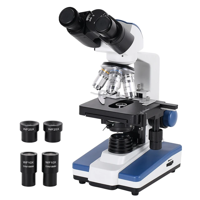
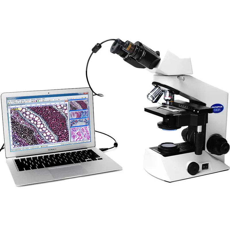
The Role of Microscopes in Unveiling Pineapple Secrets
The microscope is a pivotal tool in discovering the hidden marvels of pineapples. By magnifying the fruit’s tiniest features, it reveals secrets invisible to the naked eye. The role of microscopes in scrutinizing pineapples is profound and multifaceted.
Microscopes help us witness the precise detail of pineapple cells. With magnification, researchers can see the intricacy of cell walls and organelles. These observations lead to better understanding of the pineapple’s structure. They also unveil the processes that make up its resilient build and sweet taste.
Scientists use microscopes to study the pineapple’s fibers in great detail. They observe how these fibers intertwine, contributing to the fruit’s strength. This close view can show how fibers differ from core to surface, shedding light on growth patterns.
Microscopes also come in handy for studying pineapple enzymes like bromelain. They allow experts to identify bromelain’s presence and its distribution within the cells. This insight is vital for harnessing the enzyme’s health benefits.
Moreover, microscopes are crucial for exploring the shapes and roles of starch granules in pineapples. They make it possible to understand how the fruit stores energy. The unique appearance of these granules tells a story about the pineapple’s evolution and adaptation.
Additionally, microscopes aid in examining the skin’s defensive structures. These findings are essential for agriculture and can help protect crops from pests and diseases. Plus, they offer a glimpse into the fruit’s innate survival strategies.
Finally, researchers use microscopes to detect microscopic pathogens in pineapples. They identify harmful organisms that might affect the quality and safety of the fruit. This helps in developing measures to counter diseases and enhance pineapple cultivation.
In essence, microscopes are indispensable. They provide a detailed peek at the pineapple’s cellular life. This gives us a broader perspective and deepens our appreciation for this tropical delicacy.
 Pineapple Enzymes: A Closer Look at Bromelain
When we study ‘pineapple under a microscope’, bromelain stands out. It is a noteworthy enzyme found in pineapples. This enzyme has various health benefits, from aiding digestion to reducing inflammation.
Under the microscope, bromelain appears as tiny specks within pineapple cells. Its distribution is particularly dense in the stem and core. This pattern suggests where bromelain is most concentrated.
Scientists isolate bromelain to understand its structure and how it works. They examine how it breaks down proteins. This is why it’s commonly used as a meat tenderizer.
Medical research shows bromelain’s potential in therapeutics. Its anti-inflammatory effects may help in medical conditions like arthritis. It could also speed up healing in bruises and reduce swelling.
A microscopic view reveals the location of bromelain in the pineapple. This helps in its extraction and application in various industries. For example, it is used in dietary supplements and as a natural remedy.
Researchers use microscopes to ensure the quality of bromelain extracts. They look for purity and the correct enzyme concentration. This is important for effective use in drugs and supplements.
In summary, understanding bromelain through microscopy has real-world uses. It provides a glimpse into the enzyme’s role in health and industry. The next time we bite into a pineapple, we’re not just tasting fruit but also experiencing the effects of its powerful enzymes.
Starch Granules in Pineapple: Their Unique Appearance
Diving deeper into the microscopic world of the pineapple, we encounter its starch granules. These tiny structures are crucial in the fruit’s energy storage system. Under the microscope, the granules reveal their unique and distinct appearance.
Unlike in other plants, pineapple starch granules manifest in diverse shapes and sizes. Some take on a rounded form, while others may appear slightly oval. This variety reflects the pineapple’s complex biology and its adaptation strategies over time. The granules are densely packed within the cells, highlighting their role as energy reservoirs for the pineapple.
When ‘pineapple under a microscope’ becomes our focus, we notice the clarity of starch granules’ boundaries. The stark contrast within the cell’s interior emphasizes these structures’ significance. They are dynamic, changing their volume with the fruit’s ripening stages.
Upon the onset of ripening, the granules swell as they absorb water, an indication of the fruit’s preparation to distribute energy. As the pineapple matures, these starch reserves convert into sugars. This conversion leads to the sweetness we enjoy in a ripe pineapple.
Employing microscopes, researchers monitor these transitions closely. This enables them to decipher the precise ripening process. Beyond satisfying our taste buds, this knowledge is vital for the agriculture sector. It informs the harvesting and storage methods to ensure peak quality and flavor.
In essence, the microscopic examination of pineapple’s starch granules affords us a glimpse into the fruit’s life cycle. It evidences nature’s intricate design in shaping a pineapple’s taste and energy storage. Next time we savor a piece of pineapple, let’s ponder the microscopic transformations that have unfolded to produce its delightful sweetness.
Plant Defense Mechanisms Observed in Pineapple Skin
When delving into ‘pineapple under a microscope’, the skin of the pineapple is equally fascinating. Examining this outer layer reveals nature’s strategic defenses. Pineapple skin boasts a range of microscopic features, acting as the first line of defense against environmental threats. Here are some key observations:
- Tough Texture: The skin’s rough, tough texture is more than a tactile feature. Microscopically, it consists of thick-walled cells. These cells form a barrier, protecting the soft inner fruit from pests and harsh weather.
- Waxy Coating: A thin, waxy layer covers the skin. This coating reduces water loss and guards against fungal invasion. Dissecting this layer under the microscope shows how it repels attackers while retaining moisture.
- Specialized Cells: Looking closer, one can find specialized cells embedded in the pineapple skin. These may produce chemicals harmful to insects and fungi. These microscopic factories play a critical role in the pineapple’s survival.
Understanding these defense mechanisms helps us appreciate the pineapple’s resilience. It also provides insights into effective agricultural practices, potentially leading to more robust crop protection strategies.
 Investigating the Microscopic Pathogens in Pineapple
Studying ‘pineapple under a microscope’ also involves examining for tiny invaders. These microscopic pathogens can pose threats to the pineapple’s health. By using a microscope, scientists can spot these harmful organisms. They look for signs of diseases that could spoil the fruit or reduce its quality.
- Bacteria and Fungi: Microscopes show bacteria and fungi that may infect the pineapple. These can cause rot or disease, leading to crop losses.
- Viruses: Researchers also look for viruses at the cellular level. Some viruses can stunt pineapple growth or cause visible plant deformities.
- Parasites: Microscopes help identify parasites that feed on the pineapple. They suck nutrients, weakening the plant and affecting fruit production.
Early detection through microscopy can save entire crops. It guides farmers on when to treat or prevent infections. This knowledge is crucial for maintaining the health of pineapples.
By tackling microscopic pathogens, we can ensure the safety of pineapples. This means healthier fruit on our tables and in our stores. The next time we enjoy a pineapple, we can be grateful for the microscopic vigilance that safeguards its quality.
Understanding Pineapple’s Ripening Process on a Cellular Level
Delving into the ripening process of pineapples through the eye of a microscope is remarkable. As pineapple under a microscope becomes our study, we see cellular changes unfold. Here’s what happens on a microscopic level as pineapples ripen:
- Starch to Sugar Conversion: Initially, pineapple cells are full of starch granules. During ripening, these granules gradually transform into sugars, making the pineapple sweet.
- Cell Enlargement: Cells within the pineapple expand as the ripening process progresses. This enlargement is due to the fruit’s cells absorbing more water.
- Acid Reduction: As the pineapple ripens, the acidity in its cells starts to decrease. This change balances the fruit’s flavor, making it less tart and more enjoyable.
- Color Shift: On a cellular level, the pineapple starts to change color. This shift is from green to yellow, reflecting the degradation of chlorophyll and the production of carotenoids.
- Enzymatic Activity: Enzymes like bromelain become more active. This breaks down proteins, softening the pineapple’s texture.
The microscope is key in unveiling these subtle cellular transitions. By magnifying the pineapple cells, we gain insight into the ripening process. This knowledge helps farmers decide the best time to harvest. It also assists in optimizing storage conditions to preserve the fruit’s quality post-harvest.
In essence, a microscopic examination offers a deeper understanding of a pineapple’s journey to peak ripeness. Each stage we observe showcases nature’s precise engineering. So, when we take a bite of ripe pineapple, we taste the result of a fascinating cellular dance choreographed by the laws of biology.
 Conclusion: Unlocking the Microscopic Wonders of Pineapple
Observing a pineapple under a microscope transforms your perception of this common fruit, unveiling a world of intricate details and sophisticated structures. This exploration not only enriches your knowledge of plant biology but also bridges the gap between science and art, offering both educational and creative opportunities. By understanding the cellular architecture and unique features of pineapples, you gain a deeper appreciation for the complexity and beauty of nature. Whether you are a student, a scientist, or an art enthusiast, studying pineapples under a microscope invites you to explore the hidden wonders that lie just beneath the surface. Embrace this fascinating journey and let the microscopic world of pineapples inspire and inform your endeavors.