Introduction to Field of View in Microscopy
Understanding the field of view (FOV) in microscopy is critical. It is the extent of the observable area seen through a microscope’s eyepiece. The FOV affects how much of a specimen we can see at once. A larger field of view lets us observe more of the sample without moving it.
In microscopy, the field of view is measured in millimeters. It changes with different magnifications and microscope types. The field of view microscope becomes smaller as magnification increases. This means that while we can see finer details at higher magnifications, we see less of the sample.
It’s imperative to consider the field of view when choosing a microscope. Researchers and diagnosticians need a suitable field of view for their specific tasks. Whether counting cells or examining tissue samples, the right field of view is essential. By selecting a microscope with the appropriate field of view, these tasks become more efficient and precise.
In the next sections, we will explore how field of view varies across different microscopes, and its significance in scientific endeavors.
Importance of Field of View in Research and Diagnostics
The field of view microscope plays a pivotal role in both research and diagnostics. This is because the FOV determines the amount of a specimen visible at any one time. In research, a vast FOV allows scientists to quickly scan large sample areas. This can speed up the data collection process. It’s especially helpful when searching for rare events in a specimen, like specific cell types.
In diagnostics, the right FOV can make a significant difference. For instance, pathologists are able to detect anomalies faster when a larger portion of a tissue slide is observed. This improves accuracy in diagnoses. It also aids in identifying patterns that might indicate disease. A microscope that reveals more area at once can lead to quicker, more reliable results.
Additionally, a field of view that is too small might miss crucial details or abnormalities. This can lead to misdiagnosis or result in longer analysis times. Therefore, the impact of the FOV on outcomes cannot be overstated.
To summarize, a field of view microscope appropriate for the task can enhance efficiency and precision. It ensures that significant aspects of a specimen are not overlooked. For researchers and diagnosticians, an optimal FOV is essential for effective and accurate work.
Types of Microscopes and Their Field of View
When selecting a microscope, understanding the different types that affect the field of view is crucial.
Compound Microscopes
Compound microscopes offer high magnification for detailed inspection of tiny structures. They have a smaller field of view microscope compared to others. As magnification goes up, their FOV narrows, allowing focus on minute details but limiting the overall view.
Stereo Microscopes
Stereo microscopes, also called dissecting microscopes, provide a wider field of view. This makes them ideal for viewing larger samples in 3D. Their lower magnification means more of the sample stays visible, aiding in tasks like soldering or dissection.
Digital Microscopes
Digital microscopes combine optics with digital technology. They display the image on a monitor, often with varying FOV options. Some can manipulate the field of view through software, potentially offering a broader perspective of the specimen.
Measuring Field of View in Microscopes
To accurately gauge the field of view microscope provides, various measurement techniques are employed. These measurements are an essential aspect in selecting the right microscope for specific tasks. Here’s how the field of view is measured:
- Direct Measurement: A microscope stage micrometer, which is a slide with precise measurements marked on it, is often used. Users can directly observe and compare the known sizes on the micrometer to the field of view.
- Comparison Method: This involves observing a specimen with a known size and estimating the field of view based on its apparent size through the microscope. It’s a less precise method but can provide a quick estimate.
- Calculated Measurement: For more advanced scopes, specifications provided by manufacturers—such as the magnification and eyepiece field number—can be used to calculate the FOV. The formula usually involves dividing the eyepiece field number by the magnification factor.
- Software Assistance: Digital microscopes often come with software that can automatically calculate and display the FOV. This is especially helpful when working with variable magnifications or when comparing different magnification levels.
Understanding and measuring the field of view is of immense importance, as it influences the efficiency and accuracy with which you can observe specimens. By mastering these measurement techniques, researchers and diagnosticians can ensure they are working with a ‘field of view microscope’ that best suits their needs.
Factors Influencing Field of View
Understanding the factors that influence the field of view (FOV) in microscopy is crucial. The FOV can be affected by several components of a microscope, each playing a pivotal role in how much of a specimen is visible to the observer. Let’s explore the primary elements that dictate this all-important feature of field of view microscopes.
Objective Lenses
The objective lenses are the primary magnifying component of a microscope. They collect light from the sample and focus it to create an image. Different objectives offer varying magnifications, which directly impacts the FOV. Generally, as magnification increases, the field of view decreases. This means higher-powered objectives let you see smaller details, but less of the sample at any one time.
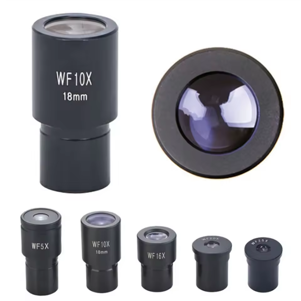
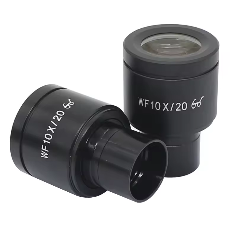
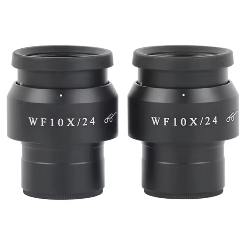
Eyepiece Lenses
Also known as oculars, eyepiece lenses are where the observer views the magnified image produced by the objective lenses. Eyepiece lenses have their own magnification factor, which can expand or narrow the FOV. Selecting eyepieces with a wider diameter generally allows for a more extensive field of view, enabling researchers to view larger areas of their sample.
Sensor Size in Digital Microscopy
In digital microscopy, the sensor size plays a significant role in determining the FOV. Larger sensors can capture more of the sample in a single image, resulting in a broader field of view. Conversely, smaller sensors might limit the visible area. When working with digital microscopes, it’s important to consider the sensor size to ensure you capture as much of the specimen as necessary for your analysis.
Enhancing Field of View with Advanced Techniques
Enhancing the field of view in microscopy can lead to better specimen analysis. Scientists and technicians can use several advanced techniques to expand the FOV. This ensures that larger sample areas are examined, improving diagnosability and research outcomes.
Panoramic Stitching
Panoramic stitching is a method that combines multiple images into one large, detailed picture. Here’s how it works in a field of view microscope:
- Capture multiple overlapping images of a sample.
- Use software to seamlessly connect these pictures.
- Create a single panoramic image that shows a larger area.
This technique allows researchers to view bigger sections of their samples than what is possible with a single image. It’s useful for mapping large specimen regions and analyzing widespread features.
Digital Zoom and Software Enhancements
Digital zoom and software play a key role in enhancing the FOV. With these tools, microscopists can:
- Enlarge specific parts of a sample without changing lenses.
- Achieve greater control over the field of view.
- Maintain image quality even when zooming digitally.
Software enhancements can also correct distortions, improve clarity, and allow for better analysis of the samples. For those using digital microscopes, these tools are vital for tailoring the field of view to specific requirements.
Practical Applications and Examples
In practical terms, the right field of view microscope can transform scientific processes. Here are some examples to illustrate the value of an appropriately sized field of view in various applications:
- Clinical Diagnostics: Pathologists rely on an expansive FOV to screen tissue samples efficiently. By seeing more of a slide at once, they detect disease markers quicker.
- Environmental Studies: Ecologists use microscopes with a wide FOV to study environmental samples, like soil or water, to monitor ecosystem health.
- Quality Control: In industries like electronics, technicians use stereo microscopes with a larger FOV to inspect circuit boards for faults.
- Education: In classrooms, a field of view microscope helps students observe a broader area, making learning more interactive and comprehensive.
- Forensic Science: Forensic scientists employ a wide FOV to examine evidence samples, ensuring nothing is missed during analysis which could be key to a case.
Each example underscores the link between the FOV and the efficiency and accuracy of the task at hand. By choosing a microscope tailored to specific needs, professionals optimize their workflow and outcomes.
Choosing the Right Microscope for Your Field of View Needs
Selecting the right field of view microscope is critical for your work’s success. To make the best choice, consider the following steps:
- Identify Your Needs: Define what you want to observe. Consider the size and detail needed.
- Understand Magnifications: Higher magnification means a smaller field of view. Decide on the balance required.
- Consider the Types: Choose between compound, stereo, or digital microscopes. Match the type to your requirements.
- Check the Eyepiece: Wider eyepieces can offer a larger field of view. Pick the one that suits your tasks.
- Evaluate the Objectives: Remember that the power of the objectives can limit your field of view.
- Assess Digital Options: If considering digital microscopes, look at sensor size and software capabilities.
- Think About Enhancements: Advanced techniques like panoramic stitching may broaden your viewing options.
- Practicality and Budget: Weigh the practical benefits against cost. Ensure the microscope delivers on both fronts.
By following these steps and understanding the elements that affect the field of view, you can choose a microscope that enhances your work’s precision and efficiency. Remember, the right field of view microscope not only improves observation but also contributes to more accurate and faster results.